 Explore
 Validate
 Learn
Learn Western blot
Antibody data
- Antibody Data
- Antigen structure
- References [3]
- Comments [0]
- Validations
- Western blot [3]
- Immunohistochemistry [1]
- Flow cytometry [1]
Submit
Validation data
Reference
Comment
Report error
- Product number
- AP7807a - Provider product page

- Provider
- Abcepta
- Proper citation
- Abgent Cat#AP7807a, RRID:AB_2242262
- Product name
- ACVRL1 Antibody (N-term)
- Antibody type
- Polyclonal
- Antigen
- Synthetic peptide
- Description
- Purified Rabbit Polyclonal Antibody (Pab)
- Reactivity
- Human, Mouse
- Host
- Rabbit
- Isotype
- IgG
- Vial size
- 400 µl
- Concentration
- 2 mg/ml
- Storage
- Maintain refrigerated at 2-8°C for up to 6 months. For long term storage store at -20°C in small aliquots to prevent freeze-thaw cycles.
Submitted references Regulation of endothelial barrier function by TGF-β type I receptor ALK5: potential role of contractile mechanisms and heat shock protein 90.
ALK1 opposes ALK5/Smad3 signaling and expression of extracellular matrix components in human chondrocytes.
LC-MS/MS analysis of apical and basolateral plasma membranes of rat renal collecting duct cells.
Antonov AS, Antonova GN, Fujii M, ten Dijke P, Handa V, Catravas JD, Verin AD
Journal of cellular physiology 2012 Feb;227(2):759-71
Journal of cellular physiology 2012 Feb;227(2):759-71
ALK1 opposes ALK5/Smad3 signaling and expression of extracellular matrix components in human chondrocytes.
Finnson KW, Parker WL, ten Dijke P, Thorikay M, Philip A
Journal of bone and mineral research : the official journal of the American Society for Bone and Mineral Research 2008 Jun;23(6):896-906
Journal of bone and mineral research : the official journal of the American Society for Bone and Mineral Research 2008 Jun;23(6):896-906
LC-MS/MS analysis of apical and basolateral plasma membranes of rat renal collecting duct cells.
Yu MJ, Pisitkun T, Wang G, Shen RF, Knepper MA
Molecular & cellular proteomics : MCP 2006 Nov;5(11):2131-45
Molecular & cellular proteomics : MCP 2006 Nov;5(11):2131-45
No comments: Submit comment
Supportive validation
- Submitted by
- Abcepta (provider)
- Main image
- Experimental details
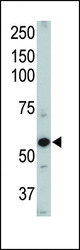
- Western blot of ACVRL1 Pab (Cat. #AP7807a). TOP LEFT: Mouse heart tissue lysate.
- Primary Ab dilution
- 1:1000

- Submitted by
- Abcepta (provider)
- Main image

- Experimental details
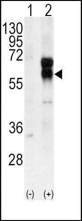
- Western blot analysis of ACVRL1 (arrow) using rabbit polyclonal ACVRL1 Antibody (N-term) (Cat. #AP7807a). 293 cell lysates (2 ug/lane) either nontransfected (Lane 1) or transiently transfected with the ACVRL1 gene (Lane 2) (Origene Technologies).
- Primary Ab dilution
- 1:1000
- Submitted by
- Abcepta (provider)
- Main image
- Experimental details
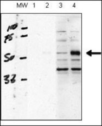
- "human chondrocytes (C28/I2 cells), transfected with empty vector (lane 1, 3) or ACVRL1(lane 2, 4). RIPA lysis buffer, 20 ug/lane of protein, primary antibody dilution 1:1000, blocking solution is 5% milk in TBST (lane 1 and 2), 5% BSA in TBST (lane 3 and 4). Data courtesy of Kenneth Finnson, Montreal General Hospital."
- Primary Ab dilution
- 1:1000

Supportive validation
- Submitted by
- Abcepta (provider)
- Main image

- Experimental details
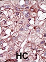
- "Formalin-fixed and paraffin-embedded human cancer tissue reacted with the primary antibody, which was peroxidase-conjugated to the secondary antibody, followed by DAB staining. This data demonstrates the use of this antibody for immunohistochemistry; clinical relevance has not been evaluated. BC = breast carcinoma; HC = hepatocarcinoma."
- Primary Ab dilution
- 1:50~100
Supportive validation
- Submitted by
- Abcepta (provider)
- Main image
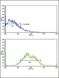
- Experimental details
- Flow cytometric analysis of HepG2 cells using ACVRL1 Antibody (N-term) (bottom histogram) compared to a negative control cell (top histogram). FITC-conjugated goat-anti-rabbit secondary antibodies were used for the analysis.
- Primary Ab dilution
- 1:10~50
