Explore
 Validate
 Learn
Learn Western blot
Antibody data
- Antibody Data
- Antigen structure
- References [0]
- Comments [0]
- Validations
- Western blot [3]
- Immunohistochemistry [1]
- Flow cytometry [1]
Submit
Validation data
Reference
Comment
Report error
- Product number
- ARG54803 - Provider product page

- Provider
- Arigo
- Product name
- anti-ACVRL1 antibody
- Antibody type
- Polyclonal
- Antigen
- KLH-conjugated synthetic peptide around aa. 38-68 (N-terminus) of Human ACVRL1.
- Description
- Purification with Protein G.
- Reactivity
- Human, Mouse
- Host
- Rabbit
- Isotype
- IgG
- Vial size
- 200 µl
- Storage
- For continuous use, store undiluted antibody at 2-8°C for up to 6 months. For long-term storage, aliquot and store at -20°C. Storage in frost free freezers is not recommended. Avoid repeated freeze/thaw cycles. The antibody solution should be gently mixed before use.
- Handling
- The antibody solution should be gently mixed before use.
No comments: Submit comment
Supportive validation
- Submitted by
- Arigo (provider)
- Main image

- Experimental details
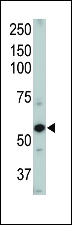
- Western blot: mouse heart tissue lysate stained with ARG54803 anti-ACVRL1 antibody.
- Submitted by
- Arigo (provider)
- Main image
- Experimental details
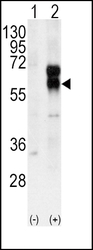
- Western blot: 2 µg of 293 cell lysates either 1) nontransfected or 2) transiently transfected with the ACVRL1 gene stained with ARG54803 anti-ACVRL1 antibody.

- Submitted by
- Arigo (provider)
- Main image
- Experimental details
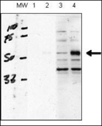
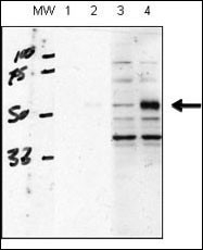
- Western blot: 20 µg of human chondrocytes (C28/I2 cells), transfected with empty vector (lane 1, 3) or ACVRL1(lane 2, 4). RIPA lysis buffer, stained with ARG54803 anti-ACVRL1 antibody at 1:1000 dilution, blocking solution is 5% milk in TBST (lane 1 and 2), 5% BSA in TBST (lane 3 and 4). Data courtesy of Kenneth Finnson, Montreal General Hospital.
Supportive validation
- Submitted by
- Arigo (provider)
- Main image
- Experimental details
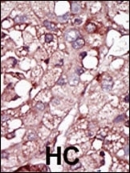
- Immunohistochemistry: formalin-fixed and paraffin-embedded human cancer tissue stained with ARG54803 anti-ACVRL1 antibody.

Supportive validation
- Submitted by
- Arigo (provider)
- Main image
- Experimental details
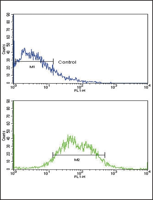
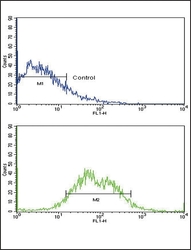
- Flow Cytometry: HepG2 cells stained with ARG54803 anti-ACVRL1 antibody (bottom histogram) or without primary antibody control (top histogram), followed by incubation with FITC labelled secondary antibody.