Explore
 Validate
 Learn
LearnHPA007042
antibody from Atlas Antibodies
Targeting: TTN
CMD1G, CMH9, CMPD4, FLJ32040, LGMD2J, MYLK5, TMD
 Immunohistochemistry
Antibody data
- Antibody Data
- Antigen structure
- References [5]
- Comments [0]
- Validations
- Immunohistochemistry [1]
Submit
Validation data
Reference
Comment
Report error
- Product number
- HPA007042 - Provider product page

- Provider
- Atlas Antibodies
- Proper citation
- Atlas Antibodies Cat#HPA007042, RRID:AB_1080292
- Product name
- Anti-TTN
- Antibody type
- Polyclonal
- Description
- Polyclonal Antibody against Human TTN, Gene description: titin, Alternative Gene Names: CMD1G, CMH9, CMPD4, FLJ32040, LGMD2J, MYLK5, TMD, Validated applications: IHC, Uniprot ID: Q8WZ42, Storage: Store at +4°C for short term storage. Long time storage is recommended at -20°C.
- Reactivity
- Human
- Host
- Rabbit
- Conjugate
- Unconjugated
- Isotype
- IgG
- Vial size
- 100 µl
- Concentration
- 0.1 mg/ml
- Storage
- Store at +4°C for short term storage. Long time storage is recommended at -20°C.
- Handling
- The antibody solution should be gently mixed before use.
Submitted references Tension-driven multi-scale self-organisation in human iPSC-derived muscle fibers
Case Report: Novel RPGRIP1L Gene Mutations Identified by Whole Exome Sequencing in a Patient With Multiple Primary Tumors
Longitudinal serum biomarker screening identifies malate dehydrogenase 2 as candidate prognostic biomarker for Duchenne muscular dystrophy
Differential Protein Expression in Congenital and Acquired Cholesteatomas
Generation and Characterization of Functional Cardiomyocytes Derived from Human T Cell-Derived Induced Pluripotent Stem Cells
Acharya A, Mao Q, Rodríguez-delaRosa A, Marchiano F, Dehapiot B, Al Tanoury Z, Rao J, Díaz-Cuadros M, Mansur A, Wagner E, Chardes C, Gupta V, Lenne P, Habermann B, Theodoly O, Pourquié O, Schnorrer F
eLife 2022;11
eLife 2022;11
Case Report: Novel RPGRIP1L Gene Mutations Identified by Whole Exome Sequencing in a Patient With Multiple Primary Tumors
Guo J, Yang Y, Ji Z, Yao M, Xia X, Sha X, Huang M
Frontiers in Genetics 2021;12
Frontiers in Genetics 2021;12
Longitudinal serum biomarker screening identifies malate dehydrogenase 2 as candidate prognostic biomarker for Duchenne muscular dystrophy
Signorelli M, Ayoglu B, Johansson C, Lochmüller H, Straub V, Muntoni F, Niks E, Tsonaka R, Persson A, Aartsma‐Rus A, Nilsson P, Al‐Khalili Szigyarto C, Spitali P
Journal of Cachexia, Sarcopenia and Muscle 2019;11(2):505-517
Journal of Cachexia, Sarcopenia and Muscle 2019;11(2):505-517
Differential Protein Expression in Congenital and Acquired Cholesteatomas
Slominski A, Shin S, Huang M, Kim S, Choi J
PLOS ONE 2015;10(9):e0137011
PLOS ONE 2015;10(9):e0137011
Generation and Characterization of Functional Cardiomyocytes Derived from Human T Cell-Derived Induced Pluripotent Stem Cells
Johnson R, Seki T, Yuasa S, Kusumoto D, Kunitomi A, Saito Y, Tohyama S, Yae K, Kishino Y, Okada M, Hashimoto H, Takei M, Egashira T, Kodaira M, Kuroda Y, Tanaka A, Okata S, Suzuki T, Murata M, Fujita J, Fukuda K
PLoS ONE 2014;9(1):e85645
PLoS ONE 2014;9(1):e85645
No comments: Submit comment
Supportive validation
- Submitted by
- Atlas Antibodies (provider)
- Enhanced method
- Orthogonal validation
- Main image
- Experimental details
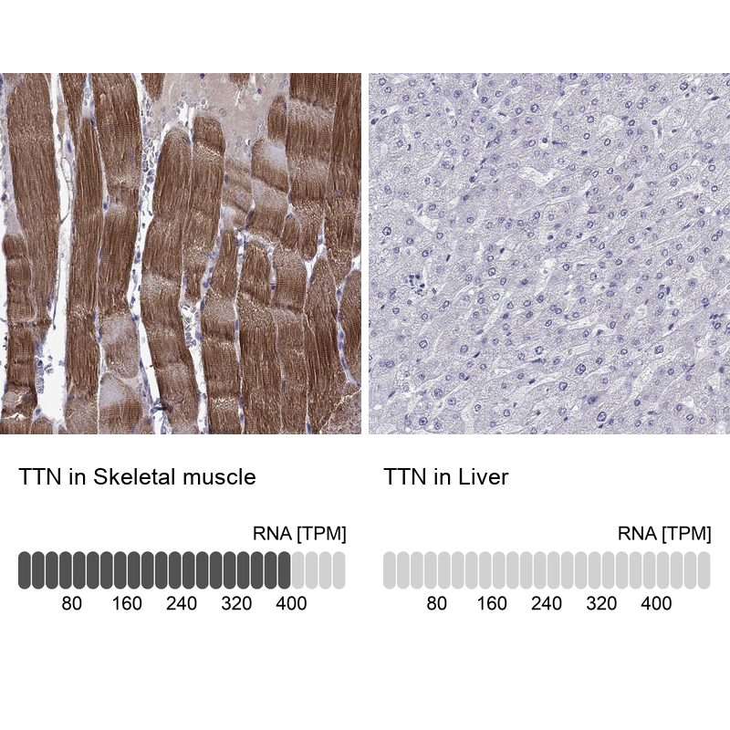
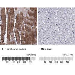
- Immunohistochemistry analysis in human skeletal muscle and liver tissues using HPA007042 antibody. Corresponding TTN RNA-seq data are presented for the same tissues.
- Sample type
- Human
- Protocol
- Protocol