Explore
 Validate
 Learn
Learn Western blot
 ELISA
ELISAAntibody data
- Antibody Data
- Antigen structure
- References [16]
- Comments [0]
- Validations
- Western blot [1]
- Immunohistochemistry [2]
Submit
Validation data
Reference
Comment
Report error
- Product number
- 13807-1-AP - Provider product page

- Provider
- Proteintech Group
- Proper citation
- Proteintech Cat#13807-1-AP, RRID:AB_2088759
- Product name
- DARS2 antibody
- Antibody type
- Polyclonal
- Description
- KD/KO validated DARS2 antibody (Cat. #13807-1-AP) is a rabbit polyclonal antibody that shows reactivity with human, mouse, rat and has been validated for the following applications: IHC, WB, ELISA.
- Reactivity
- Human, Mouse, Rat
- Host
- Rabbit
- Conjugate
- Unconjugated
- Isotype
- IgG
- Vial size
- 20ul, 150ul
Submitted references AAV9-DARS2 Gene Therapy Rescues Phenotype in Leukoencephalopathy with Brainstem and Spinal Cord Involvement and Lactate Elevation Patient Cells and Neuronal Dars2 Deficient Mice.
S-adenosylmethionine and nicotinic acid suppress Hepatocellular carcinoma proliferation by targeting SLC25A4.
DARS2 Promotes Bladder Cancer Progression by Enhancing PINK1-Mediated Mitophagy.
Secreted mitochondrial aspartyl-tRNA synthetase (DARS2) regulates TNFα signaling.
Inhibition of liver cancer cell growth by metabolites S-adenosylmethionine and nicotinic acid originating from liver progenitor cells.
eIF5A maintains intestinal epithelial homeostasis by sustaining intestinal stem cells.
Role of CENPL, DARS2, and PAICS in determining the prognosis of patients with lung adenocarcinoma.
Targeted degradation of extracellular mitochondrial aspartyl-tRNA synthetase modulates immune responses.
Mitochondrial dysfunction abrogates dietary lipid processing in enterocytes.
DARS2 is a prognostic biomarker and correlated with immune infiltrates and cuproptosis in lung adenocarcinoma.
DARS2 promotes the occurrence of lung adenocarcinoma via the ERK/c-Myc signaling pathway.
DARS2 overexpression is associated with PET/CT metabolic parameters and affects glycolytic activity in lung adenocarcinoma.
Mitochondrial metabolism coordinates stage-specific repair processes in macrophages during wound healing.
Mitochondrial oxidative phosphorylation is impaired in TALLYHO mice, a new obesity and type 2 diabetes animal model.
Loss of CLPP alleviates mitochondrial cardiomyopathy without affecting the mammalian UPRmt.
Tissue-specific loss of DARS2 activates stress responses independently of respiratory chain deficiency in the heart.
Garofolo I, Lindsay B, Liang Y, Ratajczak B, Janowski M, Walczak P, Fatemi A, Nemeth CL
Annals of neurology 2026 Jan;99(1):59-72
Annals of neurology 2026 Jan;99(1):59-72
S-adenosylmethionine and nicotinic acid suppress Hepatocellular carcinoma proliferation by targeting SLC25A4.
Liu WM, Zhang QQ, Ma HQ, Chen CY, Jiao YF, Yan HX, Yang LQ, Lai RT, He ZY
Biochemical pharmacology 2026 Jan;243(Pt 2):117530
Biochemical pharmacology 2026 Jan;243(Pt 2):117530
DARS2 Promotes Bladder Cancer Progression by Enhancing PINK1-Mediated Mitophagy.
Li D, Su H, Deng X, Huang Y, Wang Z, Zhang J, Chen C, Zheng Z, Wang Q, Zhao S, Chen ZS, Chen H, Hou L, Tan W, Li F
International journal of biological sciences 2025;21(4):1530-1544
International journal of biological sciences 2025;21(4):1530-1544
Secreted mitochondrial aspartyl-tRNA synthetase (DARS2) regulates TNFα signaling.
Johnson BS, Cornwell A, Farkas D, Yurtsever I, Joseph JA, Pradhan A, Farkas L, Londino JD, Bednash JS, Mallampalli RK
Physiological reports 2025 Nov;13(21):e70627
Physiological reports 2025 Nov;13(21):e70627
Inhibition of liver cancer cell growth by metabolites S-adenosylmethionine and nicotinic acid originating from liver progenitor cells.
Liu WM, Chen CY, Ma HQ, Zhang QQ, Zhou X, Wu YL, Huang WJ, Qi XS, Zhang YX, Tang D, Sun HY, Wu HP, Jiao YF, He ZY, Yu WF, Yan HX
Journal of gastroenterology 2025 Jun;60(6):754-769
Journal of gastroenterology 2025 Jun;60(6):754-769
eIF5A maintains intestinal epithelial homeostasis by sustaining intestinal stem cells.
Li L, Xiao Y, Liu L, Zhang Q, Zhang Y, Zhu D, Chen YG
Cell regeneration (London, England) 2025 Jun 9;14(1):23
Cell regeneration (London, England) 2025 Jun 9;14(1):23
Role of CENPL, DARS2, and PAICS in determining the prognosis of patients with lung adenocarcinoma.
Xu R, Han F, Zhao Y, Liu A, An N, Wang B, Zardo P, Sanz-Santos J, Franssen AJPM, de Loos ER, Zhao M
Translational lung cancer research 2024 Oct 31;13(10):2729-2745
Translational lung cancer research 2024 Oct 31;13(10):2729-2745
Targeted degradation of extracellular mitochondrial aspartyl-tRNA synthetase modulates immune responses.
Johnson BS, Farkas D, El-Mergawy R, Adair JA, Elhance A, Eltobgy M, Coan FM, Chafin L, Joseph JA, Cornwell A, Johns FJ, Rosas L, Rojas M, Farkas L, Bednash JS, Londino JD, Ray P, Ray A, Kagan V, Lee JS, Chen BB, Mallampalli RK
Nature communications 2024 Jul 22;15(1):6172
Nature communications 2024 Jul 22;15(1):6172
Mitochondrial dysfunction abrogates dietary lipid processing in enterocytes.
Moschandrea C, Kondylis V, Evangelakos I, Herholz M, Schneider F, Schmidt C, Yang M, Ehret S, Heine M, Jaeckstein MY, Szczepanowska K, Schwarzer R, Baumann L, Bock T, Nikitopoulou E, Brodesser S, Krüger M, Frezza C, Heeren J, Trifunovic A, Pasparakis M
Nature 2024 Jan;625(7994):385-392
Nature 2024 Jan;625(7994):385-392
DARS2 is a prognostic biomarker and correlated with immune infiltrates and cuproptosis in lung adenocarcinoma.
Liu XS, Zeng J, Zhang YH, Zhang Y, Gao Y, Liu C, Pei ZJ
American journal of cancer research 2023;13(3):818-834
American journal of cancer research 2023;13(3):818-834
DARS2 promotes the occurrence of lung adenocarcinoma via the ERK/c-Myc signaling pathway.
Fang T, Jiang J, Yu W, Li R, Tian H
Thoracic cancer 2023 Dec;14(36):3511-3521
Thoracic cancer 2023 Dec;14(36):3511-3521
DARS2 overexpression is associated with PET/CT metabolic parameters and affects glycolytic activity in lung adenocarcinoma.
Liu XS, Yuan LL, Gao Y, Ming X, Zhang YH, Zhang Y, Liu ZY, Yang Y, Pei ZJ
Journal of translational medicine 2023 Aug 26;21(1):574
Journal of translational medicine 2023 Aug 26;21(1):574
Mitochondrial metabolism coordinates stage-specific repair processes in macrophages during wound healing.
Willenborg S, Sanin DE, Jais A, Ding X, Ulas T, Nüchel J, Popović M, MacVicar T, Langer T, Schultze JL, Gerbaulet A, Roers A, Pearce EJ, Brüning JC, Trifunovic A, Eming SA
Cell metabolism 2021 Dec 7;33(12):2398-2414.e9
Cell metabolism 2021 Dec 7;33(12):2398-2414.e9
Mitochondrial oxidative phosphorylation is impaired in TALLYHO mice, a new obesity and type 2 diabetes animal model.
Hunter CA, Kartal F, Koc ZC, Murphy T, Kim JH, Denvir J, Koc EC
The international journal of biochemistry & cell biology 2019 Nov;116:105616
The international journal of biochemistry & cell biology 2019 Nov;116:105616
Loss of CLPP alleviates mitochondrial cardiomyopathy without affecting the mammalian UPRmt.
Seiferling D, Szczepanowska K, Becker C, Senft K, Hermans S, Maiti P, König T, Kukat A, Trifunovic A
EMBO reports 2016 Jul;17(7):953-64
EMBO reports 2016 Jul;17(7):953-64
Tissue-specific loss of DARS2 activates stress responses independently of respiratory chain deficiency in the heart.
Dogan SA, Pujol C, Maiti P, Kukat A, Wang S, Hermans S, Senft K, Wibom R, Rugarli EI, Trifunovic A
Cell metabolism 2014 Mar 4;19(3):458-69
Cell metabolism 2014 Mar 4;19(3):458-69
No comments: Submit comment
Supportive validation
- Submitted by
- Proteintech Group (provider)
- Main image

- Experimental details
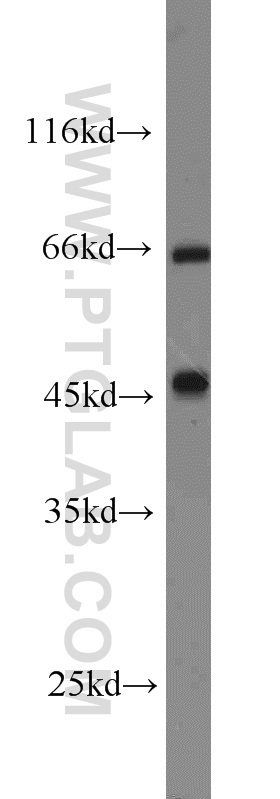
- K-562 cells were subjected to SDS PAGE followed by western blot with 13807-1-AP(DARS2 antibody) at dilution of 1:800
- Sample type
- cell line
Supportive validation
- Submitted by
- Proteintech Group (provider)
- Main image

- Experimental details
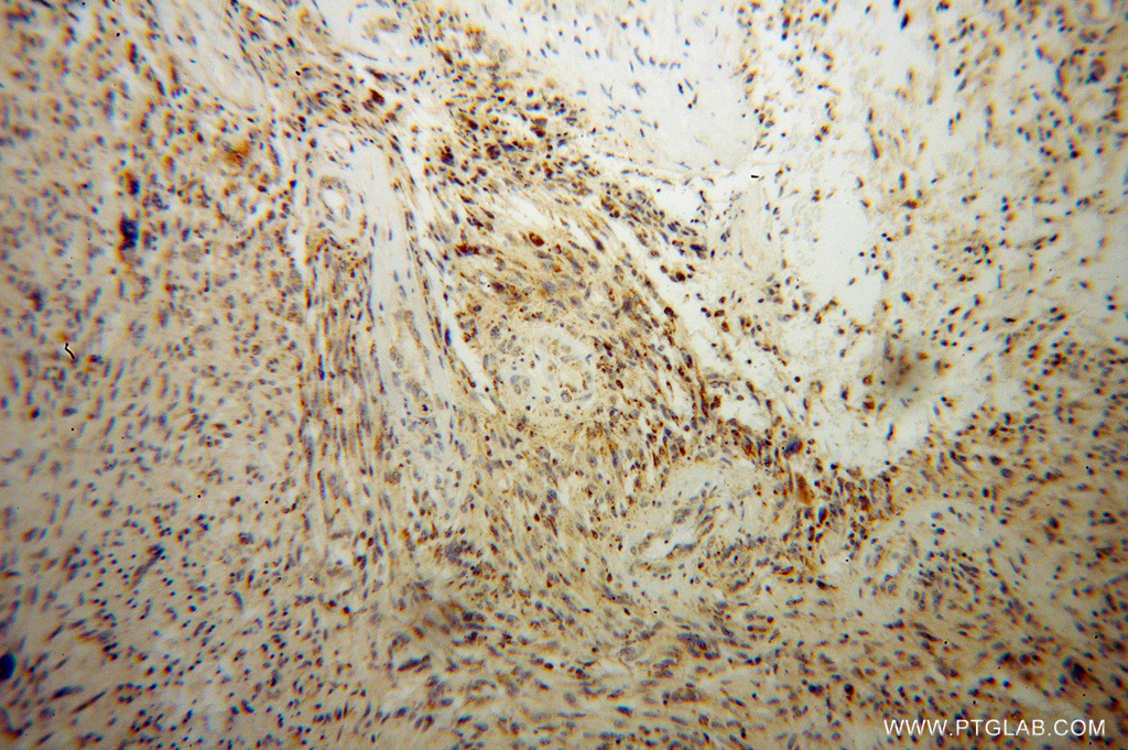
- Immunohistochemical of paraffin-embedded human gliomas using 13807-1-AP(DARS2 antibody) at dilution of 1:100 (under 10x lens)
- Sample type
- tissue
- Submitted by
- Proteintech Group (provider)
- Main image
- Experimental details
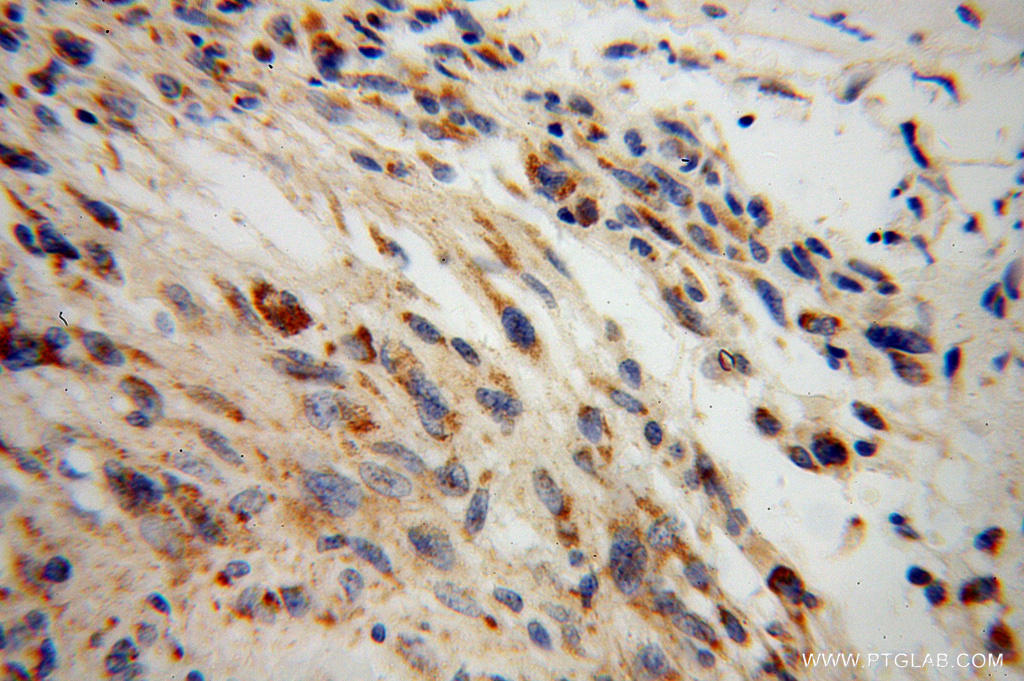
- The DARS2 antibody from Proteintech is a rabbit polyclonal antibody to a recombinant protein of human DARS2. This antibody recognizes human,mouse,rat antigen. The DARS2 antibody has been validated for the following applications: ELISA, WB, IHC analysis.