Explore
 Validate
 Learn
Learn Western blot
Antibody data
- Antibody Data
- Antigen structure
- References [1]
- Comments [0]
- Validations
- Western blot [3]
- Immunohistochemistry [1]
Submit
Validation data
Reference
Comment
Report error
- Product number
- AP8141a - Provider product page

- Provider
- Abcepta
- Proper citation
- Abgent Cat#AP8141a, RRID:AB_2279615
- Product name
- HK1 (Hexokinase) Antibody (N-term)
- Antibody type
- Polyclonal
- Antigen
- Synthetic peptide
- Description
- Purified Rabbit Polyclonal Antibody (Pab)
- Reactivity
- Human, Mouse
- Host
- Rabbit
- Isotype
- IgG
- Vial size
- 400 µl
- Concentration
- 0.35 mg/ml
- Storage
- Maintain refrigerated at 2-8°C for up to 6 months. For long term storage store at -20°C in small aliquots to prevent freeze-thaw cycles.
Submitted references Quantitative changes in the mitochondrial proteome from subjects with mild cognitive impairment, early stage, and late stage Alzheimer's disease.
Lynn BC, Wang J, Markesbery WR, Lovell MA
Journal of Alzheimer's disease : JAD 2010;19(1):325-39
Journal of Alzheimer's disease : JAD 2010;19(1):325-39
No comments: Submit comment
Supportive validation
- Submitted by
- Abcepta (provider)
- Main image

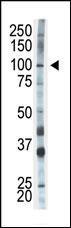
- Experimental details
- The anti-HK1 Pab (Cat. #AP8141a) is used in Western blot to detect HK1 in mouse skeletal muscle tissue lysate.
- Primary Ab dilution
- 1:1000
- Submitted by
- Abcepta (provider)
- Main image
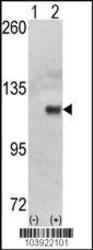
- Experimental details
- Western blot analysis of HK1 (arrow) using HK1 Antibody (N-term) (Cat.#AP8141a). 293 cell lysates (2 ug/lane) either nontransfected (Lane 1) or transiently transfected with the HK1 gene (Lane 2) (Origene Technologies).
- Primary Ab dilution
- 1:1000

- Submitted by
- Abcepta (provider)
- Main image

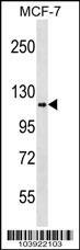
- Experimental details
- HK1 Antibody (L93) (Cat. #AP8141a) western blot analysis in MCF-7 cell line lysates (35ug/lane).This demonstrates the HK1 antibody detected the HK1 protein (arrow).
- Primary Ab dilution
- 1:1000
Supportive validation
- Submitted by
- Abcepta (provider)
- Main image
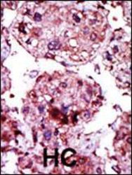
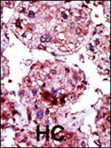
- Experimental details
- "Formalin-fixed and paraffin-embedded human cancer tissue reacted with the primary antibody, which was peroxidase-conjugated to the secondary antibody, followed by AEC staining. This data demonstrates the use of this antibody for immunohistochemistry; clinical relevance has not been evaluated. BC = breast carcinoma; HC = hepatocarcinoma."
- Primary Ab dilution
- 1:50~100