Explore
 Validate
 Learn
Learn Western blot
 ELISA
ELISAAntibody data
- Antibody Data
- Antigen structure
- References [0]
- Comments [0]
- Validations
- Western blot [5]
Submit
Validation data
Reference
Comment
Report error
- Product number
- NBP1-04295 - Provider product page

- Provider
- Novus Biologicals
- Proper citation
- Novus Cat#NBP1-04295, RRID:AB_1521169
- Product name
- Mouse Monoclonal Hexokinase 1 Antibody
- Antibody type
- Monoclonal
- Description
- Protein G purified. The antibody recognizes all four isoforms of Hexokinase (1~4) in recombinant protein.
- Reactivity
- Human
- Host
- Mouse
- Isotype
- IgG
- Vial size
- 0.1 ml
- Concentration
- 1.0 mg/ml
- Storage
- Store at 4C short term. Aliquot and store at -20C long term. Avoid freeze-thaw cycles.
No comments: Submit comment
Supportive validation
- Submitted by
- Novus Biologicals (provider)
- Main image
- Experimental details
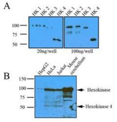
- Western Blot: Hexokinase 1 Antibody (4D7) [NBP1-04295] - (A): Recombinant protein (20ng or 100ng) of Hexokinase four isoform were resolved by SDS-PAGE, transferred to NC membrane and probed with anti-human Hexokinase (1:1000). (B): Cell lysates (20ug) were resolved by SDS-PAGE, transferred to NC membrane and probed with anti-human Hexokinase (1:1,000). Proteins were visualized using a goat anti-mouse secondary antibody conjugated to HRP and an ECL detection system.

- Submitted by
- Novus Biologicals (provider)
- Main image
- Experimental details
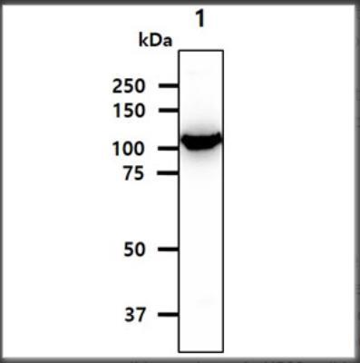
- Western Blot: Hexokinase 1 Antibody (4D7) [NBP1-04295] - Cell lysates of HepG2 (30ug) were resolved by SDS-PAGE, transferred to PVDF membrane and probed with anti-human Hexokinase1 (1:1000). Proteins were visualized using a goat anti-mouse secondary antibody conjugated to HRP and an ECL detection.
- Submitted by
- Novus Biologicals (provider)
- Main image
- Experimental details
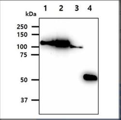
- Western Blot: Hexokinase 1 Antibody (4D7) [NBP1-04295] - The Recombinant Proteins (20ng) were resolved by SDS-PAGE, transferred to PVDF membrane and probed with anti-human Hexokinase antibody (1:1000). Proteins were visualized using a goat anti-mouse secondary antibody conjugated to HRP and an ECL detection system. Lane 1.: Hexokinase 1 recombinant protein Lane 2.: Hexokinase 2 recombinant protein Lane 3.: Hexokinase 3 recombinant protein Lane 4.: Hexokinase 4 recombinant protein

- Submitted by
- Novus Biologicals (provider)
- Main image
- Experimental details
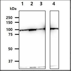
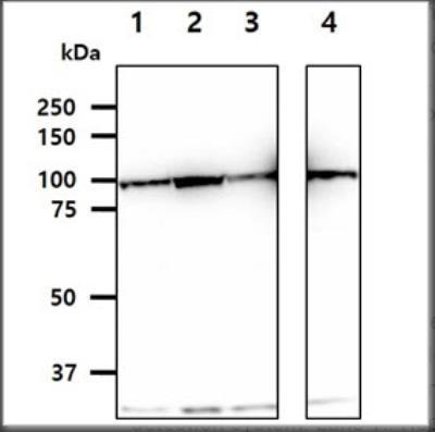
- Western Blot: Hexokinase 1 Antibody (4D7) [NBP1-04295] - The cell lysates (40ug) were resolved by SDS-PAGE, transferred to PVDF membrane and probed with anti-human Hexokinase antibody (1:1000). Proteins were visualized using a goat anti-mouse secondary antibody conjugated to HRP and an ECL detection system. Lane 1.: HepG2 cell lysate Lane 2.: HeLa cell lysate Lane 3.: Jurkat cell lysate Lane 4.: K562 cell lysate
- Submitted by
- Novus Biologicals (provider)
- Main image
- Experimental details
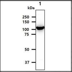
- Western Blot: Hexokinase 1 Antibody (4D7) [NBP1-04295] - The tissue lysate (40ug) were resolved by SDS-PAGE, transferred to PVDF membrane and probed with anti-human Hexokinase antibody (1:1000). Proteins were visualized using a goat anti-mouse secondary antibody conjugated to HRP and an ECL detection system. Lane 1.: Brain tissue lysate