Explore
 Validate
 Learn
Learn Western blot
 Immunohistochemistry
Antibody data
- Antibody Data
- Antigen structure
- References [3]
- Comments [0]
- Validations
- Immunohistochemistry [1]
Submit
Validation data
Reference
Comment
Report error
- Product number
- MAB1624 - Provider product page

- Provider
- Novus Biologicals
- Product name
- Mouse Monoclonal Tenascin R Antibody
- Antibody type
- Monoclonal
- Description
- Protein A or G purified from hybridoma culture supernatant. Detects mouse and rat Tenascin R in Western blots.
- Reactivity
- Mouse, Rat
- Host
- Mouse
- Isotype
- IgG
- Vial size
- 100 ug
- Concentration
- LYOPH
- Storage
- Use a manual defrost freezer and avoid repeated freeze-thaw cycles. 12 months from date of receipt, -20 to -70 degreesC as supplied. 1 month, 2 to 8 degreesC under sterile conditions after reconstitution. 6 months, -20 to -70 degreesC under sterile conditions after reconstitution.
Submitted references The altered expression of perineuronal net elements during neural differentiation.
Quantitative Visualization of Dynamic Tracer Transportation in the Extracellular Space of Deep Brain Regions Using Tracer-Based Magnetic Resonance Imaging.
Quantitative Visualization of Dynamic Tracer Transportation in the Extracellular Space of Deep Brain Regions Using Tracer-Based Magnetic Resonance Imaging.
Eskici NF, Erdem-Ozdamar S, Dayangac-Erden D
Cellular & molecular biology letters 2018;23:5
Cellular & molecular biology letters 2018;23:5
Quantitative Visualization of Dynamic Tracer Transportation in the Extracellular Space of Deep Brain Regions Using Tracer-Based Magnetic Resonance Imaging.
Hou J, Wang W, Quan X, Liang W, Li Z, Chen D, Han H
Medical science monitor : international medical journal of experimental and clinical research 2017 Sep 3;23:4260-4268
Medical science monitor : international medical journal of experimental and clinical research 2017 Sep 3;23:4260-4268
No comments: Submit comment
Supportive validation
- Submitted by
- Novus Biologicals (provider)
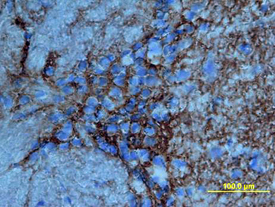
- Main image

- Experimental details
- Tenascin R in Rat Brain . Tenascin R was detected in perfusion fixed frozen sections of rat brain using Mouse Anti-Mouse/Rat Tenascin R Monoclonal Antibody (Catalog # MAB1624) at 1 µg/mL overnight at 4 °C. Tissue was stained using the Anti-Mouse HRP-DAB Cell & Tissue Staining Kit (brown; Catalog # CTS002) and counterstained with hematoxylin (blue). View our protocol for Chromogenic IHC Staining of Frozen Tissue Sections.