Explore
 Validate
 Learn
Learn Western blot
 Immunohistochemistry
Antibody data
- Antibody Data
- Antigen structure
- References [2]
- Comments [0]
- Validations
- Immunohistochemistry [1]
Submit
Validation data
Reference
Comment
Report error
- Product number
- AF3865 - Provider product page

- Provider
- R&D Systems
- Product name
- Human Tenascin R Antibody
- Antibody type
- Polyclonal
- Description
- Immunogen affinity purified. Detects human Tenascin R in direct ELISAs and Western blots. In direct ELISAs and Western blots, less than 1% cross-reactivity with recombinant human Tenascin C is observed.
- Reactivity
- Human
- Host
- Goat
- Conjugate
- Unconjugated
- Antigen sequence
Q92752- Isotype
- IgG
- Vial size
- 100 ug
- Concentration
- LYOPH
- Storage
- Use a manual defrost freezer and avoid repeated freeze-thaw cycles. 12 months from date of receipt, -20 to -70 °C as supplied. 1 month, 2 to 8 °C under sterile conditions after reconstitution. 6 months, -20 to -70 °C under sterile conditions after reconstitution.
Submitted references Sensory experience-dependent formation of perineuronal nets and expression of Cat-315 immunoreactive components in the mouse somatosensory cortex.
Extracellular matrix molecules exhibit unique expression pattern in the climbing fiber-generating precerebellar nucleus, the inferior olive.
Ueno H, Suemitsu S, Okamoto M, Matsumoto Y, Ishihara T
Neuroscience 2017 Jul 4;355:161-174
Neuroscience 2017 Jul 4;355:161-174
Extracellular matrix molecules exhibit unique expression pattern in the climbing fiber-generating precerebellar nucleus, the inferior olive.
Kecskes S, Gaál B, Rácz É, Birinyi A, Hunyadi A, Matesz C
Neuroscience 2015 Jan 22;284:412-21
Neuroscience 2015 Jan 22;284:412-21
No comments: Submit comment
Supportive validation
- Submitted by
- R&D Systems (provider)
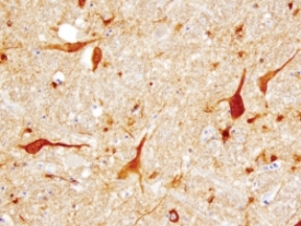
- Main image
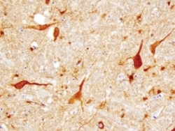
- Experimental details
- Tenascin R in Human Brainstem. Tenascin R was detected in immersion fixed paraffin-embedded sections of human brainstem (medulla) using 1.7 µg/mL Goat Anti-Human Tenascin R Antigen Affinity-purified Polyclonal Antibody (Catalog # AF3865) overnight at 4 °C. Tissue was stained with the Anti-Goat HRP-DAB Cell & Tissue Staining Kit (brown; Catalog # CTS008) and counterstained with hematoxylin (blue). View our protocol for Chromogenic IHC Staining of Paraffin-embedded Tissue Sections.