Explore
 Validate
 Learn
Learn Immunocytochemistry
Antibody data
- Antibody Data
- Antigen structure
- References [8]
- Comments [0]
- Validations
- Immunocytochemistry [1]
- Immunohistochemistry [1]
- Flow cytometry [1]
Submit
Validation data
Reference
Comment
Report error
- Product number
- MAB1448 - Provider product page

- Provider
- R&D Systems
- Product name
- Human Alkaline Phosphatase/ALPL Antibody
- Antibody type
- Monoclonal
- Description
- Protein A or G purified from hybridoma culture supernatant. Detects liver, bone and kidney Alkaline Phosphatase/ALPL from human tissue (2).
- Reactivity
- Human
- Host
- Mouse
- Conjugate
- Unconjugated
- Isotype
- IgG
- Antibody clone number
- B4-78
- Vial size
- 100 ug
- Concentration
- LYOPH
- Storage
- Use a manual defrost freezer and avoid repeated freeze-thaw cycles. 12 months from date of receipt, -20 to -70 °C as supplied. 1 month, 2 to 8 °C under sterile conditions after reconstitution. 6 months, -20 to -70 °C under sterile conditions after reconstitution.
Submitted references Growth Factor Screening in Dystrophic Muscles Reveals PDGFB/PDGFRB-Mediated Migration of Interstitial Stem Cells.
V-myc immortalizes human neural stem cells in the absence of pluripotency-associated traits.
Osteodifferentiated mesenchymal stem cells from bone marrow and adipose tissue express HLA-G and display immunomodulatory properties in HLA-mismatched settings: implications in bone repair therapy.
Cementum- and periodontal ligament-like tissue formation by dental follicle cell sheets co-cultured with Hertwig's epithelial root sheath cells.
In vivo impact of a 4 bp deletion mutation in the DLX3 gene on bone development.
Substance P stimulates bone marrow stromal cell osteogenic activity, osteoclast differentiation, and resorption activity in vitro.
The influence of proepicardial cells on the osteogenic potential of marrow stromal cells in a three-dimensional tubular scaffold.
Bone marrow-derived osteoblast progenitor cells in circulating blood contribute to ectopic bone formation in mice.
Camps J, Grosemans H, Gijsbers R, Maes C, Sampaolesi M
International journal of molecular sciences 2019 Mar 5;20(5)
International journal of molecular sciences 2019 Mar 5;20(5)
V-myc immortalizes human neural stem cells in the absence of pluripotency-associated traits.
Pino-Barrio MJ, García-García E, Menéndez P, Martínez-Serrano A
PloS one 2015;10(3):e0118499
PloS one 2015;10(3):e0118499
Osteodifferentiated mesenchymal stem cells from bone marrow and adipose tissue express HLA-G and display immunomodulatory properties in HLA-mismatched settings: implications in bone repair therapy.
Montespan F, Deschaseaux F, Sensébé L, Carosella ED, Rouas-Freiss N
Journal of immunology research 2014;2014:230346
Journal of immunology research 2014;2014:230346
Cementum- and periodontal ligament-like tissue formation by dental follicle cell sheets co-cultured with Hertwig's epithelial root sheath cells.
Bai Y, Bai Y, Matsuzaka K, Hashimoto S, Fukuyama T, Wu L, Miwa T, Liu X, Wang X, Inoue T
Bone 2011 Jun 1;48(6):1417-26
Bone 2011 Jun 1;48(6):1417-26
In vivo impact of a 4 bp deletion mutation in the DLX3 gene on bone development.
Choi SJ, Roodman GD, Feng JQ, Song IS, Amin K, Hart PS, Wright JT, Haruyama N, Hart TC
Developmental biology 2009 Jan 1;325(1):129-37
Developmental biology 2009 Jan 1;325(1):129-37
Substance P stimulates bone marrow stromal cell osteogenic activity, osteoclast differentiation, and resorption activity in vitro.
Wang L, Zhao R, Shi X, Wei T, Halloran BP, Clark DJ, Jacobs CR, Kingery WS
Bone 2009 Aug;45(2):309-20
Bone 2009 Aug;45(2):309-20
The influence of proepicardial cells on the osteogenic potential of marrow stromal cells in a three-dimensional tubular scaffold.
Valarmathi MT, Yost MJ, Goodwin RL, Potts JD
Biomaterials 2008 May;29(14):2203-16
Biomaterials 2008 May;29(14):2203-16
Bone marrow-derived osteoblast progenitor cells in circulating blood contribute to ectopic bone formation in mice.
Otsuru S, Tamai K, Yamazaki T, Yoshikawa H, Kaneda Y
Biochemical and biophysical research communications 2007 Mar 9;354(2):453-8
Biochemical and biophysical research communications 2007 Mar 9;354(2):453-8
No comments: Submit comment
Supportive validation
- Submitted by
- R&D Systems (provider)
- Main image
- Experimental details
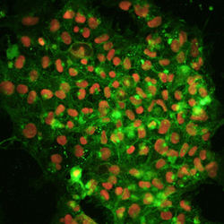
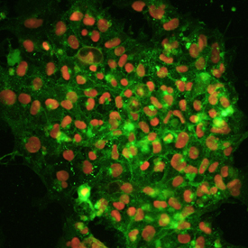
- Alkaline Phosphatase and Oct-3/4 in BG01V Human Stem Cells. Alkaline Phosphatase/ALPL and Oct-3/4 were detected in human BG01V embryonic stem cells using 10 µg/mL Mouse Anti-Human Alkaline Phosphatase/ALPL Monoclonal Antibody (Catalog # MAB1448) and 10 µg/mL Human Oct-3/4 Antigen Affinity-purified Polyclonal Antibody (Catalog # AF1759). Cells were incubated with primary antibodies for 3 hours at room temperature. Cells were stained for Alkaline Phosphatase/ALPL using the NorthernLights™ 557-conjugated Anti-Mouse IgG Secondary Antibody (pseudo-stained green; Catalog # NL007), and stained for Oct-3/4 using the NorthernLights 637-conjugated Anti-Goat IgG Secondary Antibody (red; Catalog # NL002). View our protocol for Fluorescent ICC Staining of Cells on Coverslips.
Supportive validation
- Submitted by
- R&D Systems (provider)
- Main image
- Experimental details
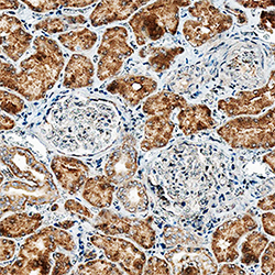
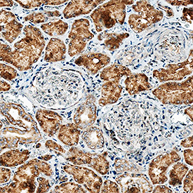
- Alkaline Phosphatase/ALPL in Human Kidney. Alkaline Phosphatase/ALPL was detected in immersion fixed paraffin-embedded sections of human kidney using Mouse Anti-Human Alkaline Phosphatase/ALPL Monoclonal Antibody (Catalog # MAB1448) at 15 µg/mL overnight at 4 °C. Tissue was stained using the Anti-Mouse HRP-DAB Cell & Tissue Staining Kit (brown; Catalog # CTS002) and counterstained with hematoxylin (blue). Specific staining was localized to cytoplasm in epithelial cells. View our protocol for Chromogenic IHC Staining of Paraffin-embedded Tissue Sections.
Supportive validation
- Submitted by
- R&D Systems (provider)
- Main image
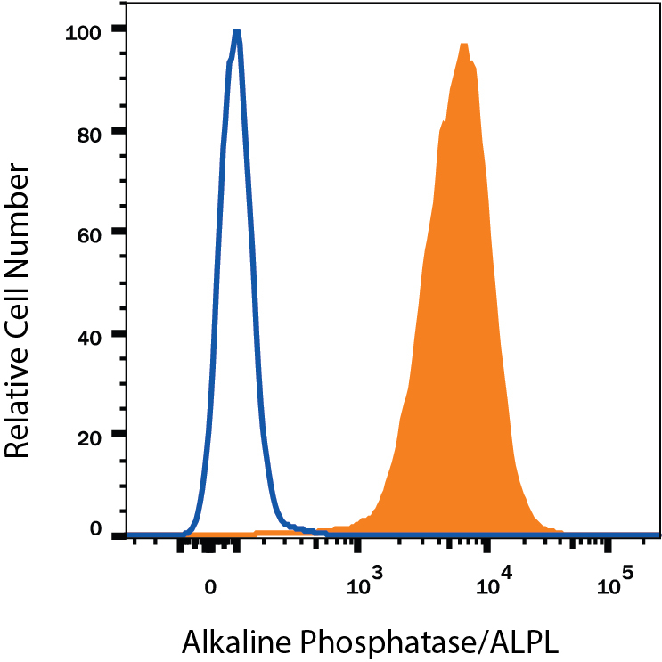
- Experimental details
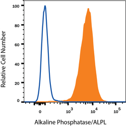
- Detection of Alkaline Phosphatase/ALPL in BG01V Human Cells by Flow Cytometry. BG01V human embryonic stem cells was stained with Mouse Anti-Human Alkaline Phosphatase/ALPL Monoclonal Antibody (Catalog # MAB1448, filled histogram) or isotype control antibody (Catalog # MAB002, open histogram), followed by Phycoerythrin-conjugated Anti-Mouse IgG Secondary Antibody (Catalog # F0102B).