Explore
 Validate
 Learn
Learn ELISA
ELISA Other assay
Antibody data
- Antibody Data
- Antigen structure
- References [1]
- Comments [0]
- Validations
- Other assay [5]
Submit
Validation data
Reference
Comment
Report error
- Product number
- PA5-72930 - Provider product page

- Provider
- Invitrogen Antibodies
- Product name
- NKIAMRE Polyclonal Antibody
- Antibody type
- Polyclonal
- Antigen
- Synthetic peptide
- Reactivity
- Human
- Host
- Rabbit
- Isotype
- IgG
- Vial size
- 100 μL
- Concentration
- 1 mg/mL
- Storage
- Store at 4°C short term. For long term storage, store at -20°C, avoiding freeze/thaw cycles.
Submitted references CDKL3 promotes osteosarcoma progression by activating Akt/PKB.
He A, Ma L, Huang Y, Zhang H, Duan W, Li Z, Fei T, Yuan J, Wu H, Liu L, Bai Y, Dai W, Wang Y, Li H, Sun Y, Wang Y, Wang C, Yuan T, Yang Q, Tian S, Dong M, Sheng R, Xiang D
Life science alliance 2020 May;3(5)
Life science alliance 2020 May;3(5)
No comments: Submit comment
Supportive validation
- Submitted by
- Invitrogen Antibodies (provider)
- Main image
- Experimental details
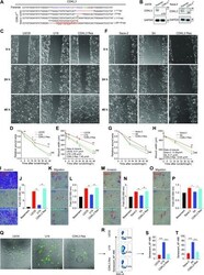
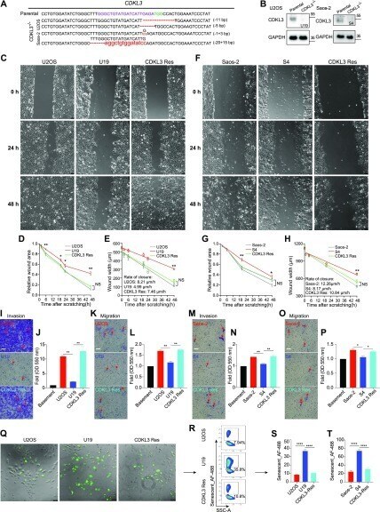
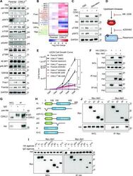
- Figure 2. Knockdown of cyclin-dependent kinase-like 3 (CDKL3) inhibited osteosarcoma (OS) cell invasion and migration. (A) Genomic DNA sequencing of CDKL3-KO clones. (B) CDKL3 expression in CDKL3-KO and parental cells by Western blot analysis. (C) Microscopy images of wound closure of parental, CDKL3-KO (U19), and CDKL3-rescued (Res) U2OS cells at 0, 24, and 48 h after scratching. Scale bar = 75 mum. (D) Quantification of the wounded area invaded during 48 h of U2OS cells (n = 3). (E) Quantification of wound healing speed (mum/h) of U2OS cells (n = 3). (F) Microscopy images of wound closure of parental, CDKL3-KO (S4), and CDKL3-rescued (Res) Saos-2 cells at 0, 24, and 48 h after scratching. Scale bar = 75 mum. (G) Quantification of the wounded area invaded during 48 h of Saos-2 cells (n = 3). (H) Quantification of wound healing speed (mum/h) of Saos-2 cells (n = 3). (I) Representative images of Transwell-invaded U2OS cells (purple stained, red arrows). Scale bar = 100 mum. (J) Quantification of stained/invaded cells in (I) (n = 3). (K) Representative images of Transwell-migrated U2OS cells (purple stained, red arrows). Scale bar = 100 mum. (L) Quantification of stained/migrated cells in (K) (n = 3). (M) Representative images of Transwell-invaded Saos-2 cells (purple stained, red arrows). Scale bar = 100 mum. (N) Quantification of stained/invaded cells in (M) (n = 3). (O) Representative images of Transwell-migrated Saos-2 cells (purple stained, red arrows). Scale bar = 100 mum
- Submitted by
- Invitrogen Antibodies (provider)
- Main image
- Experimental details
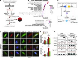
- Figure 4. Cyclin-dependent kinase-like 3 (CDKL3) inhibits autophagy in osteosarcoma (OS). (A) An outline of OS clinical dataset analysis (GEO accession: GSE21257 ). 33 out of 53 patient samples were selectively analyzed based on the CDKL3 expression level for the differential gene expression patterns. (B) Signaling pathway was mapped by the R package clusterProfiler. Upper and bottom figures represent down-regulated and up-regulated pathways (CDKL3 top 0.3 versus bottom 0.3), respectively. KEGG enrichment analysis using the clusterProfiler R package was performed on differential expressed genes with a strict cutoff of P < 0.01 and false discovery rate of less than 0.05. The size of the dot indicates the number of differentially expressed genes in the pathway. Color of the dot represents the value of Benjamini and Hochberg false discovery rate-adjusted P -value. (C) Molecular connection of PI3K-Akt and AMPK pathway in regulation of cell growth, autophagy, etc. This summarized mechanism was extracted from the GEO dataset GSE21257 . mTORC1 plays key roles in eukaryotic cell metabolism by promoting cell growth and inhibiting autophagy and apoptosis. (D) Representative images of parental cells and CDKL3-KO U2OS and Saos-2 cells transfected with mRFP-eGFP-LC3, at normal conditions (DMEM with 10% FBS, control) or 6 h EBSS treatment (starvation). Colocalized mRFP and eGFP (yellow dots) indicate autophagosomes, only mRFP (red dots) indicates autolysosomes. Nuclei (blue) were labeled w

- Submitted by
- Invitrogen Antibodies (provider)
- Main image
- Experimental details
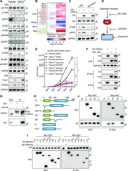
- Figure 5. Cyclin-dependent kinase-like 3 (CDKL3) critically regulates Akt activation in osteosarcoma (OS). (A) Akt and mTOR activation under regular or starvation conditions in parental and CDKL3-KO U2OS cells. Upon CDKL3 KO, Akt and mTORC1 activation were significantly alleviated. The numbers below each blotting strip is intensity quantification by ImageJ. (B) The expression patterns of mTORC1 and FoxO downstream target genes confirm that CDKL3 regulates both pathways at the transcription level. Cells were cultured under the regular growth condition. (C) Overexpression of CDKL3 in U2OS cells causes hyper-activation of Akt in the presence and absence of FBS. (D) General working mechanism of rapamycin, AZD5363, and MK-2206. (E) U2OS cell growth under different conditions (n = 3). (F) Co-immunoprecipitation (Co-IP) reveals the physical interaction between HA-CDKL3 and Myc-Akt1. (G) Endogenous CDKL3 coexists with endogenous Akt shown by co-IP. (H) Schematic diagrams of Akt1 constructs for mapping. (I) Co-IP of HA-CDKL3 with different Akt1 constructs shows that the Akt kinase domain is indispensable for CDKL3 interaction. (I, J) Reverse IP of CDKL3 and Akt1 confirms the findings in (I). Error bars indicate SD (n = 3). * P < 0.05 parental DMSO versus CDKL3 -/- DMSO, or parental rapamycin, or parental AZD5363, or parental MK-2206; ** P < 0.01 CDKL3 -/- DMSO versus CDKL3 -/- rapamycin, or CDKL3 -/- AZD5363, or CDKL3 -/- MK-2206; ## P < 0.01 parental MK-2206 versus CDKL3 -/- MK-2206.
- Submitted by
- Invitrogen Antibodies (provider)
- Main image
- Experimental details
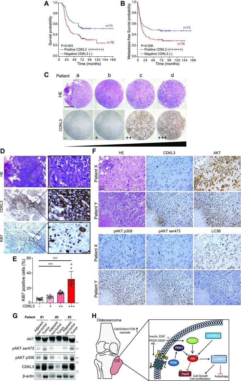
- Figure 6. Cyclin-dependent kinase-like 3 (CDKL3) defines poor prognosis and correlates with Akt phosphorylation in clinic. (A, B) Kaplan-Meier plots of overall survival (A) and metastasis-free survival (B) of 152 osteosarcoma (OS) patients, stratified by CDKL3 levels (- represents negative staining; +, ++, and +++ represent weak, intermediate, and strong staining, respectively). (C) Representative immunohistochemistry (IHC) images of OS biopsies with different levels of CDKL3 expression on an OS microarray containing 152 primary OS tissues samples. Scale bar = 500 mum. (D) Representative HE and IHC images of OS biopsies stained by CDKL3 and Ki67. Scale bar = 100 mum. (E) Quantitative analysis of Ki67 expression in OS patients with different levels of CDKL3 expression. (F) HE and IHC staining images of CDKL3, AKT, pAKT (p308), pAKT (ser473), and LC3 in representative CDKL3-positive and CDKL3-negative patients. Scale bar = 100 mum. (G) Western blot detection of CDKL3 and Akt phosphorylation in OS tissues and adjacent non-tumor tissues from three different patients. (H) Putative underlying mechanism that CDKL3 promotes OS progression. Overexpression of CDKL3 leads to increased phosphorylation of Akt, followed by governing mTORC1 and FoxO activities, likely independent of functions of PDK1, growth factors, or relevant receptors; this may inhibit autophagy and eventually promote OS development. * P < 0.05, ** P < 0.01, *** P < 0.001, two-tailed t test. Source data are available fo
- Submitted by
- Invitrogen Antibodies (provider)
- Main image
- Experimental details
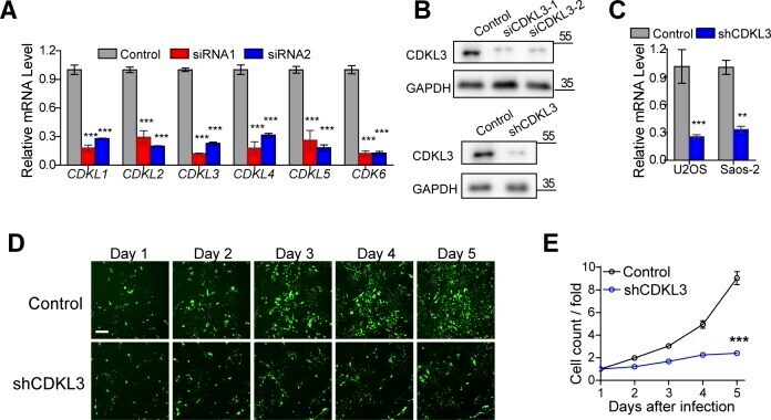
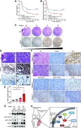
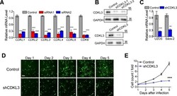
- Figure S2. Knockdown of cyclin-dependent kinase-like 3 (CDKL3) inhibited osteosarcoma (OS) cell growth. (A) qRT-PCR analysis in U2OS cells transfected with siRNAs. Error bars indicate SD (n = 3). (B) Western blotting confirming the knockdown of CDKL3 in U2OS cells. (C) qRT-PCR analysis in targeted cells transfected with shRNAs. Error bars indicate SD (n = 5). (D) Fluorescence photomicrographs of Saos-2 cells in consecutive 5 d after shCDKL3 infection at a magnification of 100x. Scale bar = 150 mum. (E) Fold change of cell count in consecutive 5 d, which equals to cell counts divided by the cell counts in the first day after infection. Control: Saos-2 cells infected with lentivirus containing control shRNA. shCDKL3: Saos-2 cells infected with lentivirus specific interfering of CDKL3 (n = 3). Error bars indicate SD (n = 3 or n = 5). *** P < 0.001, two-tailed t test. Source data are available for this figure. Source Data for Figure S2