Explore
 Validate
 Learn
Learn Western blot
 Immunocytochemistry
Antibody data
- Antibody Data
- Antigen structure
- References [2]
- Comments [0]
- Validations
- Immunocytochemistry [1]
- Chromatin Immunoprecipitation [1]
Submit
Validation data
Reference
Comment
Report error
- Product number
- HPA051228 - Provider product page

- Provider
- Atlas Antibodies
- Proper citation
- Atlas Antibodies Cat#HPA051228, RRID:AB_2681396
- Product name
- Anti-ZNF281
- Antibody type
- Polyclonal
- Description
- Polyclonal Antibody against Human ZNF281, Gene description: zinc finger protein 281, Alternative Gene Names: ZBP-99, Validated applications: WB, ICC, ChIP, Uniprot ID: Q9Y2X9, Storage: Store at +4°C for short term storage. Long time storage is recommended at -20°C.
- Reactivity
- Human
- Host
- Rabbit
- Conjugate
- Unconjugated
- Isotype
- IgG
- Vial size
- 100 µl
- Concentration
- 0.1 mg/ml
- Storage
- Store at +4°C for short term storage. Long time storage is recommended at -20°C.
- Handling
- The antibody solution should be gently mixed before use.
Submitted references ZNF281/Zfp281 is a target of miR‐1 and counteracts muscle differentiation
Systematic assessment of cervical cancer initiation and progression uncovers genetic panels for deep learning-based early diagnosis and proposes novel diagnostic and prognostic biomarkers.
Nicolai S, Pieraccioli M, Smirnov A, Pitolli C, Anemona L, Mauriello A, Candi E, Annicchiarico‐Petruzzelli M, Shi Y, Wang Y, Melino G, Raschellà G
Molecular Oncology 2019;14(2):294-308
Molecular Oncology 2019;14(2):294-308
Systematic assessment of cervical cancer initiation and progression uncovers genetic panels for deep learning-based early diagnosis and proposes novel diagnostic and prognostic biomarkers.
Long NP, Jung KH, Yoon SJ, Anh NH, Nghi TD, Kang YP, Yan HH, Min JE, Hong SS, Kwon SW
Oncotarget 2017 Dec 12;8(65):109436-109456
Oncotarget 2017 Dec 12;8(65):109436-109456
No comments: Submit comment
Supportive validation
- Submitted by
- Atlas Antibodies (provider)
- Main image
- Experimental details
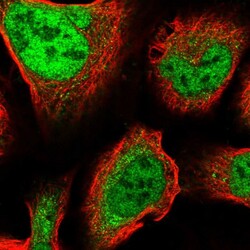
- Immunofluorescent staining of human cell line U-2 OS shows localization to nucleoplasm.
- Sample type
- Human

Supportive validation
- Submitted by
- Atlas Antibodies (provider)
- Main image
- Experimental details
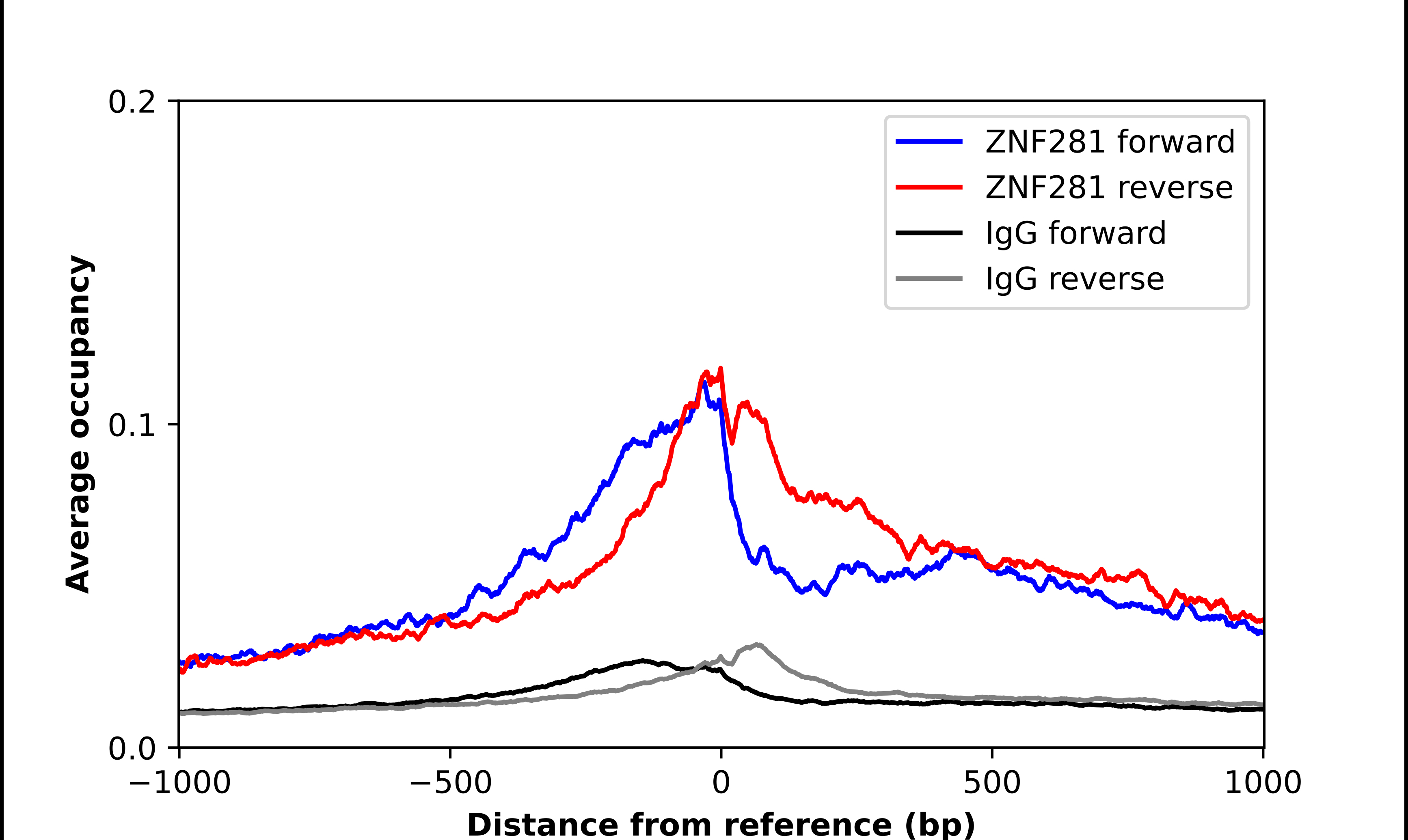
- ChIP-Exo-Seq composite graph for Anti-ZNF281 (HPA051228, Lot 000016842) tested in K562 cells. Strand-specific reads (blue: forward, red: reverse) and IgG controls (black: forward, grey: reverse) are plotted against the distance from a composite set of reference binding sites. The antibody exhibits robust target enrichment compared to a non-specific IgG control and precisely reveals its structural organization around the binding site. Data generated by Prof. B. F. Pugh´s Lab at Cornell University.