Explore
 Validate
 Learn
Learn Western blot
 Immunocytochemistry
 Immunoprecipitation
Antibody data
- Antibody Data
- Antigen structure
- References [5]
- Comments [0]
- Validations
- Immunocytochemistry [1]
- Other assay [1]
Submit
Validation data
Reference
Comment
Report error
- Product number
- 32-5700 - Provider product page

- Provider
- Invitrogen Antibodies
- Product name
- PKP1 Monoclonal Antibody (10B2)
- Antibody type
- Monoclonal
- Antigen
- Other
- Reactivity
- Human
- Host
- Mouse
- Isotype
- IgG
- Antibody clone number
- 10B2
- Vial size
- 100 μg
- Concentration
- 0.5 mg/mL
- Storage
- -20°C
Submitted references Analysis of buccal mucosa as a prognostic tool in children with arrhythmogenic cardiomyopathy.
Homozygous splice site mutations in PKP1 result in loss of epidermal plakophilin 1 expression and underlie ectodermal dysplasia/skin fragility syndrome in two consanguineous families.
Defining desmosomal plakophilin-3 interactions.
Desmoglein 4 in hair follicle differentiation and epidermal adhesion: evidence from inherited hypotrichosis and acquired pemphigus vulgaris.
Desmoglein 4 in hair follicle differentiation and epidermal adhesion: evidence from inherited hypotrichosis and acquired pemphigus vulgaris.
Bueno-Beti C, Field E, Tsatsopoulou A, Perry G, Sheppard MN, Behr ER, Saffitz JE, Kaski JP, Asimaki A
Progress in pediatric cardiology 2022 Mar;64:None
Progress in pediatric cardiology 2022 Mar;64:None
Homozygous splice site mutations in PKP1 result in loss of epidermal plakophilin 1 expression and underlie ectodermal dysplasia/skin fragility syndrome in two consanguineous families.
Sprecher E, Molho-Pessach V, Ingber A, Sagi E, Indelman M, Bergman R
The Journal of investigative dermatology 2004 Mar;122(3):647-51
The Journal of investigative dermatology 2004 Mar;122(3):647-51
Defining desmosomal plakophilin-3 interactions.
Bonné S, Gilbert B, Hatzfeld M, Chen X, Green KJ, van Roy F
The Journal of cell biology 2003 Apr 28;161(2):403-16
The Journal of cell biology 2003 Apr 28;161(2):403-16
Desmoglein 4 in hair follicle differentiation and epidermal adhesion: evidence from inherited hypotrichosis and acquired pemphigus vulgaris.
Kljuic A, Bazzi H, Sundberg JP, Martinez-Mir A, O'Shaughnessy R, Mahoney MG, Levy M, Montagutelli X, Ahmad W, Aita VM, Gordon D, Uitto J, Whiting D, Ott J, Fischer S, Gilliam TC, Jahoda CA, Morris RJ, Panteleyev AA, Nguyen VT, Christiano AM
Cell 2003 Apr 18;113(2):249-60
Cell 2003 Apr 18;113(2):249-60
No comments: Submit comment
Supportive validation
- Submitted by
- Invitrogen Antibodies (provider)
- Main image
- Experimental details
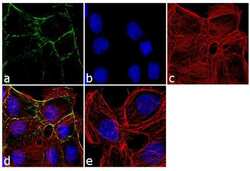
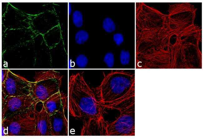
- Immunofluorescence analysis of Plakophilin (PKP1) was performed using 70% confluent log phase A-431 cells. The cells were fixed with 4% paraformaldehyde for 10 minutes, permeabilized with 0.1% Triton X-100 for 10 minutes, and blocked with 1% BSA for 1 hour at room temperature. The cells were labeled with PKP1 (10B2) Mouse Monoclonal Antibody (Product # 32-5700) at 2 µg/mL in 0.1% BSA and incubated for 3 hours at room temperature and then labeled with Goat anti-Mouse IgG (H+L) Superclonal Secondary Antibody, Alexa Fluor® 488 conjugate (Product # A28175) at a dilution of 1:2000 for 45 minutes at room temperature (Panel a: green). Nuclei (Panel b: blue) were stained with SlowFade® Gold Antifade Mountant with DAPI (Product # S36938). F-actin (Panel c: red) was stained with Alexa Fluor® 555 Rhodamine Phalloidin (Product # R415, 1:300). Panel d represents the merged image showing cell junctional localization. Panel e shows the no primary antibody control. The images were captured at 60X magnification.
Supportive validation
- Submitted by
- Invitrogen Antibodies (provider)
- Main image
- Experimental details
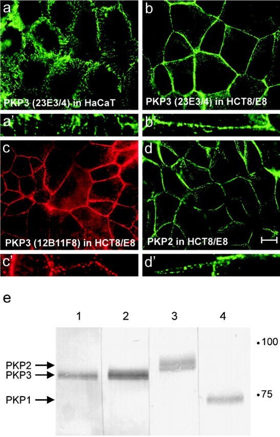
- Figure 1. Immunodetection of PKPs using different mouse mAbs. PKP3, detected with mAb 23E3/4 (a and b), 12B11F8 (c), or PKP2 (d) show a similar expression pattern along cell-cell contacts in methanol-fixed HaCaT (a) and HCT8/E8 (b-d) cells. Larger magnifications display the punctate localization of these proteins along cell-cell contacts (a'-d'). Bar, 10 mum. (e) Western blot detection of PKP3 (lane 1, mAb 12B11F8; lane 2, 23E3/4), PKP2 (lane 3), and PKP1 (lane 4) in HaCaT protein lysates. Equal amounts of protein were loaded in each lane.