Explore
 Validate
 Learn
Learn Western blot
 Immunohistochemistry
Antibody data
- Antibody Data
- Antigen structure
- References [3]
- Comments [0]
- Validations
- Immunohistochemistry [1]
Submit
Validation data
Reference
Comment
Report error
- Product number
- HPA028732 - Provider product page

- Provider
- Atlas Antibodies
- Proper citation
- Atlas Antibodies Cat#HPA028732, RRID:AB_10602916
- Product name
- Anti-LAD1
- Antibody type
- Polyclonal
- Description
- Polyclonal Antibody against Human LAD1, Gene description: ladinin 1, Validated applications: WB, IHC, Uniprot ID: O00515, Storage: Store at +4°C for short term storage. Long time storage is recommended at -20°C.
- Reactivity
- Human
- Host
- Rabbit
- Conjugate
- Unconjugated
- Isotype
- IgG
- Vial size
- 100 µl
- Concentration
- 0.1 mg/ml
- Storage
- Store at +4°C for short term storage. Long time storage is recommended at -20°C.
- Handling
- The antibody solution should be gently mixed before use.
Submitted references Ladinin-1 in actin arcs of oral squamous cell carcinoma is involved in cell migration and epithelial phenotype
Biomarkers Found in the Tumor Interstitial Fluid may Help Explain the Differential Behavior Among Keratinocyte Carcinomas
SILAC identifies LAD1 as a filamin-binding regulator of actin dynamics in response to EGF and a marker of aggressive breast tumors
Abé T, Yamazaki M, Nozumi M, Maruyama S, Takamura K, Ohashi R, Ajioka Y, Tanuma J
Scientific Reports 2024;14(1)
Scientific Reports 2024;14(1)
Biomarkers Found in the Tumor Interstitial Fluid may Help Explain the Differential Behavior Among Keratinocyte Carcinomas
Matas-Nadal C, Bech-Serra J, Gatius S, Gomez X, Ribes-Santolaria M, Guasch-Vallés M, Pedraza N, Casanova J, de la Torre Gómez C, Garí E, Aguayo-Ortiz R
Molecular & Cellular Proteomics 2023;22(6):100547
Molecular & Cellular Proteomics 2023;22(6):100547
SILAC identifies LAD1 as a filamin-binding regulator of actin dynamics in response to EGF and a marker of aggressive breast tumors
Roth L, Srivastava S, Lindzen M, Sas-Chen A, Sheffer M, Lauriola M, Enuka Y, Noronha A, Mancini M, Lavi S, Tarcic G, Pines G, Nevo N, Heyman O, Ziv T, Rueda O, Gnocchi D, Pikarsky E, Admon A, Caldas C, Yarden Y
Science Signaling 2018;11(515)
Science Signaling 2018;11(515)
No comments: Submit comment
Supportive validation
- Submitted by
- Atlas Antibodies (provider)
- Enhanced method
- Orthogonal validation
- Main image
- Experimental details
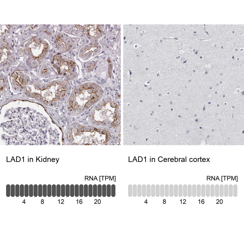
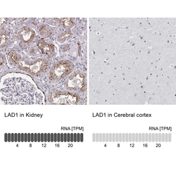
- Immunohistochemistry analysis in human kidney and cerebral cortex tissues using HPA028732 antibody. Corresponding LAD1 RNA-seq data are presented for the same tissues.
- Sample type
- Human
- Protocol
- Protocol