Explore
 Validate
 Learn
Learn Western blot
 Immunocytochemistry
Antibody data
- Antibody Data
- Antigen structure
- References [2]
- Comments [0]
- Validations
- Immunocytochemistry [1]
- Immunohistochemistry [1]
Submit
Validation data
Reference
Comment
Report error
- Product number
- AMAb90860 - Provider product page

- Provider
- Atlas Antibodies
- Proper citation
- Atlas Antibodies Cat#AMAb90860, RRID:AB_2665696
- Product name
- Anti-KDM5B
- Antibody type
- Monoclonal
- Description
- Monoclonal Antibody against Human KDM5B, Clone ID: CL1129, Gene description: lysine (K)-specific demethylase 5B, Alternative Gene Names: CT31, JARID1B, PLU-1, RBBP2H1A, Validated applications: ICC, WB, IHC, Uniprot ID: Q9UGL1, Storage: Store at +4°C for short term storage. Long time storage is recommended at -20°C.
- Reactivity
- Human
- Host
- Mouse
- Conjugate
- Unconjugated
- Isotype
- IgG
- Antibody clone number
- CL1129
- Vial size
- 100 µl
- Concentration
- 1.0 mg/ml
- Storage
- Store at +4°C for short term storage. Long time storage is recommended at -20°C.
- Handling
- The antibody solution should be gently mixed before use.
Submitted references Androgen Effects on Alcohol-induced Liver Fibrosis Are Controlled by a Notch-dependent Epigenetic Switch
Profiling cancer testis antigens in non-small-cell lung cancer.
Nataraj K, Schonfeld M, Rodriguez A, Sharma M, Weinman S, Tikhanovich I
Cellular and Molecular Gastroenterology and Hepatology 2025;19(1):101414
Cellular and Molecular Gastroenterology and Hepatology 2025;19(1):101414
Profiling cancer testis antigens in non-small-cell lung cancer.
Djureinovic D, Hallström BM, Horie M, Mattsson JSM, La Fleur L, Fagerberg L, Brunnström H, Lindskog C, Madjar K, Rahnenführer J, Ekman S, Ståhle E, Koyi H, Brandén E, Edlund K, Hengstler JG, Lambe M, Saito A, Botling J, Pontén F, Uhlén M, Micke P
JCI insight 2016 Jul 7;1(10):e86837
JCI insight 2016 Jul 7;1(10):e86837
No comments: Submit comment
Supportive validation
- Submitted by
- Atlas Antibodies (provider)
- Main image
- Experimental details
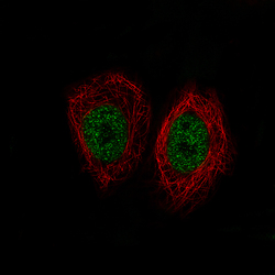
- Immunofluorescence staining of MCF7 cells using the Anti-KDM5B monoclonal antibody, showing specific staining in the nucleoplasm in green. Microtubule- and nuclear probes are visualized in red and blue, respectively (where available).
- Sample type
- Human

Supportive validation
- Submitted by
- Atlas Antibodies (provider)
- Enhanced method
- Orthogonal validation
- Main image
- Experimental details
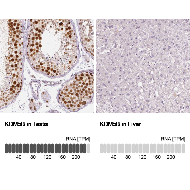
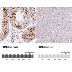
- Immunohistochemistry analysis in human testis and liver tissues using AMAb90860 antibody. Corresponding KDM5B RNA-seq data are presented for the same tissues.
- Sample type
- Human
- Protocol
- Protocol