Explore
 Validate
 Learn
Learn Western blot
 Immunocytochemistry
 Gel shift
Antibody data
- Antibody Data
- Antigen structure
- References [11]
- Comments [0]
- Validations
- Immunocytochemistry [4]
- Immunoprecipitation [1]
- Immunohistochemistry [1]
- Other assay [4]
Submit
Validation data
Reference
Comment
Report error
- Product number
- MA5-11486 - Provider product page

- Provider
- Invitrogen Antibodies
- Product name
- Myogenin Monoclonal Antibody (F5D)
- Antibody type
- Monoclonal
- Antigen
- Recombinant full-length protein
- Description
- MA5-11486 targets Myogenin in GS, IF, IHC (P), IP, and WB applications and shows reactivity with Feline, Human, mouse, and Rat samples. The MA5-11486 immunogen is recombinant protein containing rat myogenin aa 30-224.
- Reactivity
- Human, Mouse, Rat, Feline
- Host
- Mouse
- Isotype
- IgG
- Antibody clone number
- F5D
- Vial size
- 500 μL
- Concentration
- 0.5 mg/mL
- Storage
- 4°C
Submitted references RNA-Seq analysis of a Pax3-expressing myoblast clone in-vitro and effect of culture surface stiffness on differentiation.
Dysregulation of Muscle-Specific MicroRNAs as Common Pathogenic Feature Associated with Muscle Atrophy in ALS, SMA and SBMA: Evidence from Animal Models and Human Patients.
HSP70 drives myoblast fusion during C2C12 myogenic differentiation.
Recovery of fertility in azoospermia rats after injection of adipose-tissue-derived mesenchymal stem cells: the sperm generation.
A comprehensive characterization study of human bone marrow mscs with an emphasis on molecular and ultrastructural properties.
Mesenchymal stem cells improve the healing of ischemic colonic anastomoses (experimental study).
Molecular characterization of an EWSR1-POU5F1 fusion associated with a t(6;22) in an undifferentiated soft tissue sarcoma.
Endometriosis-associated skeletal muscle regeneration: a hitherto undescribed entity and a potential diagnostic pitfall.
Isolation and in vitro characterisation of dental pulp stem cells from natal teeth.
Characterization of mesenchymal stem cells from rat bone marrow: ultrastructural properties, differentiation potential and immunophenotypic markers.
Pulmonary meningothelial-like nodules: new insights into a common but poorly understood entity.
Richardson L, Wang D, Hughes R, Johnson CA, Peckham M
Scientific reports 2022 Feb 18;12(1):2841
Scientific reports 2022 Feb 18;12(1):2841
Dysregulation of Muscle-Specific MicroRNAs as Common Pathogenic Feature Associated with Muscle Atrophy in ALS, SMA and SBMA: Evidence from Animal Models and Human Patients.
Malacarne C, Galbiati M, Giagnorio E, Cavalcante P, Salerno F, Andreetta F, Cagnoli C, Taiana M, Nizzardo M, Corti S, Pensato V, Venerando A, Gellera C, Fenu S, Pareyson D, Masson R, Maggi L, Dalla Bella E, Lauria G, Mantegazza R, Bernasconi P, Poletti A, Bonanno S, Marcuzzo S
International journal of molecular sciences 2021 May 26;22(11)
International journal of molecular sciences 2021 May 26;22(11)
HSP70 drives myoblast fusion during C2C12 myogenic differentiation.
Thakur SS, Swiderski K, Chhen VL, James JL, Cranna NJ, Islam AMT, Ryall JG, Lynch GS
Biology open 2020 Jul 22;9(7)
Biology open 2020 Jul 22;9(7)
Recovery of fertility in azoospermia rats after injection of adipose-tissue-derived mesenchymal stem cells: the sperm generation.
Cakici C, Buyrukcu B, Duruksu G, Haliloglu AH, Aksoy A, Isık A, Uludag O, Ustun H, Subası C, Karaoz E
BioMed research international 2013;2013:529589
BioMed research international 2013;2013:529589
A comprehensive characterization study of human bone marrow mscs with an emphasis on molecular and ultrastructural properties.
Karaöz E, Okçu A, Gacar G, Sağlam O, Yürüker S, Kenar H
Journal of cellular physiology 2011 May;226(5):1367-82
Journal of cellular physiology 2011 May;226(5):1367-82
Mesenchymal stem cells improve the healing of ischemic colonic anastomoses (experimental study).
Adas G, Arikan S, Karatepe O, Kemik O, Ayhan S, Karaoz E, Kamali G, Eryasar B, Ustek D
Langenbeck's archives of surgery 2011 Jan;396(1):115-26
Langenbeck's archives of surgery 2011 Jan;396(1):115-26
Molecular characterization of an EWSR1-POU5F1 fusion associated with a t(6;22) in an undifferentiated soft tissue sarcoma.
Deng FM, Galvan K, de la Roza G, Zhang S, Souid AK, Stein CK
Cancer genetics 2011 Aug;204(8):423-9
Cancer genetics 2011 Aug;204(8):423-9
Endometriosis-associated skeletal muscle regeneration: a hitherto undescribed entity and a potential diagnostic pitfall.
Colella R, Mameli MG, Bellezza G, Sordo RD, Cavaliere A, Sidoni A
The American journal of surgical pathology 2010 Jan;34(1):10-7
The American journal of surgical pathology 2010 Jan;34(1):10-7
Isolation and in vitro characterisation of dental pulp stem cells from natal teeth.
Karaöz E, Doğan BN, Aksoy A, Gacar G, Akyüz S, Ayhan S, Genç ZS, Yürüker S, Duruksu G, Demircan PC, Sariboyaci AE
Histochemistry and cell biology 2010 Jan;133(1):95-112
Histochemistry and cell biology 2010 Jan;133(1):95-112
Characterization of mesenchymal stem cells from rat bone marrow: ultrastructural properties, differentiation potential and immunophenotypic markers.
Karaoz E, Aksoy A, Ayhan S, Sariboyaci AE, Kaymaz F, Kasap M
Histochemistry and cell biology 2009 Nov;132(5):533-46
Histochemistry and cell biology 2009 Nov;132(5):533-46
Pulmonary meningothelial-like nodules: new insights into a common but poorly understood entity.
Mukhopadhyay S, El-Zammar OA, Katzenstein AL
The American journal of surgical pathology 2009 Apr;33(4):487-95
The American journal of surgical pathology 2009 Apr;33(4):487-95
No comments: Submit comment
Supportive validation
- Submitted by
- Invitrogen Antibodies (provider)
- Main image

- Experimental details
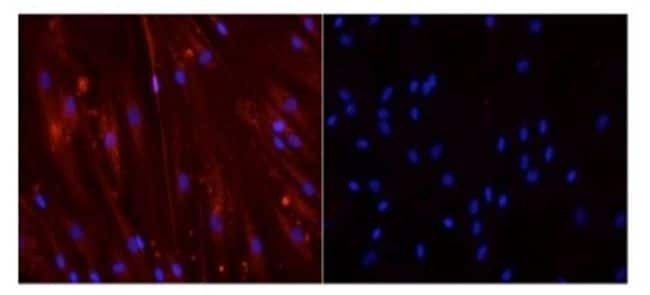
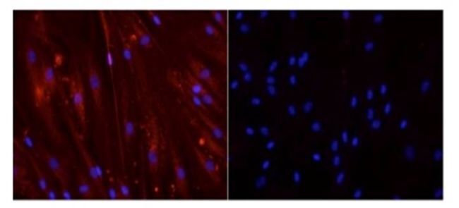
- Human skeletal muscle myoblasts (hSMM) fixed in 3.7% formaldehyde and permeabilized with 0.2% Triton X-100 were stained with either Myogenin Monoclonal Antibody (Product # MA5-11486) at 1:10 dilution and an anti-mouse Texas Red secondary antibody (left) or secondary antibody alone (right). Nuclei were stained with DAPI (blue). Data courtesy of Innovators Program.
- Submitted by
- Invitrogen Antibodies (provider)
- Main image
- Experimental details
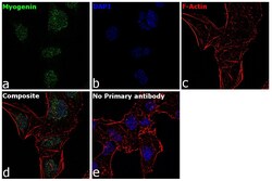
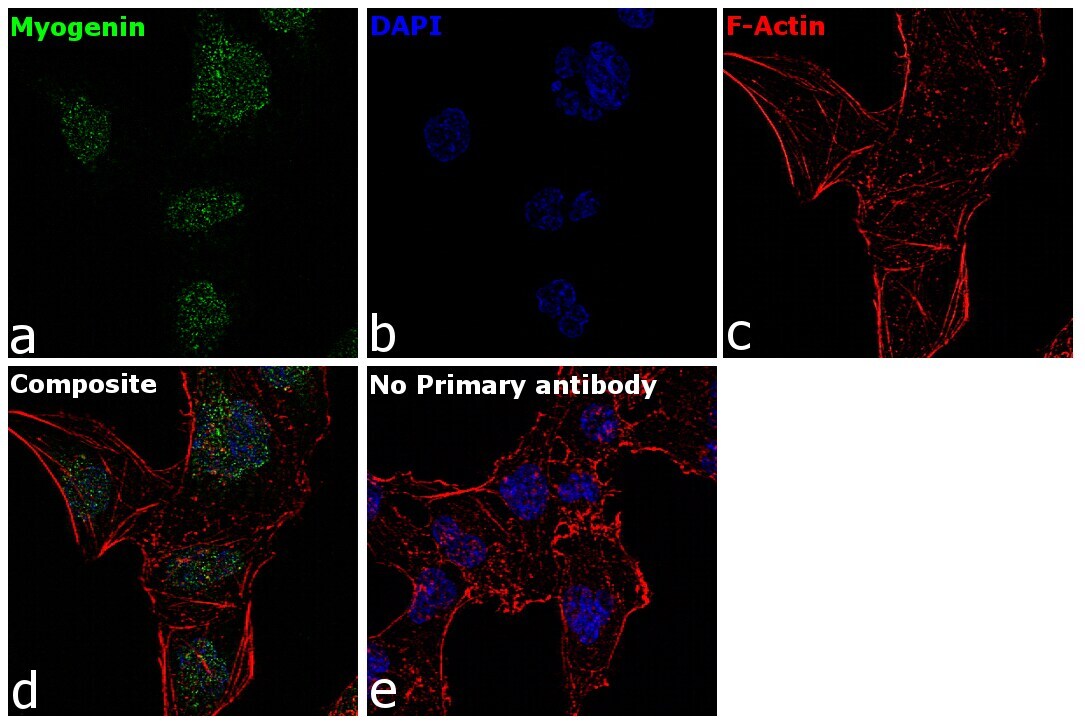
- Immunofluorescence analysis of Myogenin was performed using 70% confluent log phase RD cells. The cells were fixed with 4% paraformaldehyde for 10 minutes, permeabilized with 0.1% Triton™ X-100 for 15 minutes, and blocked with 2% BSA for 1 hour at room temperature. The cells were labeled with Myogenin Mouse Monoclonal Antibody (F5D) Product # MA5-11486) at 5 µg/mL in 0.1% BSA, incubated at 4 degree Celsius overnight and then with Goat anti-Mouse IgG (H+L) Superclonal™ Recombinant Secondary Antibody, Alexa Fluor® 488 conjugate (Product # A28175) at a dilution of 1:2000 for 45 minutes at room temperature (Panel a: green). Nuclei (Panel b: blue) were stained with ProLong™ Diamond Antifade Mountant with DAPI (Product # P36962). F-actin (Panel c: red) was stained with Rhodamine Phalloidin (Product # R415, 1:300). Panel d represents the merged image showing nuclear localization. Panel e represents control cells with no primary antibody to assess background. The images were captured at 60X magnification.

- Submitted by
- Invitrogen Antibodies (provider)
- Main image
- Experimental details
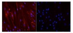
- Human skeletal muscle myoblasts (hSMM) fixed in 3.7% formaldehyde and permeabilized with 0.2% Triton X-100 were stained with either Myogenin Monoclonal Antibody (Product # MA5-11486) at 1:10 dilution and an anti-mouse Texas Red secondary antibody (left) or secondary antibody alone (right). Nuclei were stained with DAPI (blue). Data courtesy of Innovators Program.
- Submitted by
- Invitrogen Antibodies (provider)
- Main image

- Experimental details
- Immunofluorescence analysis of Myogenin was performed using 70% confluent log phase RD cells. The cells were fixed with 4% paraformaldehyde for 10 minutes, permeabilized with 0.1% Triton™ X-100 for 15 minutes, and blocked with 2% BSA for 1 hour at room temperature. The cells were labeled with Myogenin Mouse Monoclonal Antibody (F5D) Product # MA5-11486) at 5 µg/mL in 0.1% BSA, incubated at 4 degree Celsius overnight and then with Goat anti-Mouse IgG (H+L) Superclonal™ Recombinant Secondary Antibody, Alexa Fluor® 488 conjugate (Product # A28175) at a dilution of 1:2000 for 45 minutes at room temperature (Panel a: green). Nuclei (Panel b: blue) were stained with ProLong™ Diamond Antifade Mountant with DAPI (Product # P36962). F-actin (Panel c: red) was stained with Rhodamine Phalloidin (Product # R415, 1:300). Panel d represents the merged image showing nuclear localization. Panel e represents control cells with no primary antibody to assess background. The images were captured at 60X magnification.
Supportive validation
- Submitted by
- Invitrogen Antibodies (provider)
- Main image
- Experimental details
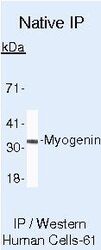
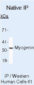
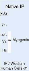
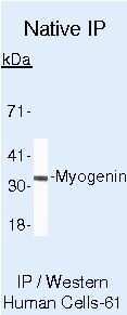
- Immunoprecipitation of Myogenin using Myogenin Monoclonal Antibody (Product # MA5-11486) on Native Human Rh30 Cells.
Supportive validation
- Submitted by
- Invitrogen Antibodies (provider)
- Main image
- Experimental details
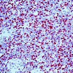
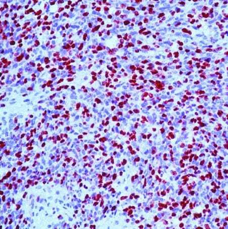
- Formalin-fixed, paraffin-embedded human rhabdomyosarcoma stained with Myogenin antibody using peroxidase-conjugate and AEC chromogen. Note nuclear staining of tumor cells.
Supportive validation
- Submitted by
- Invitrogen Antibodies (provider)
- Main image
- Experimental details
- Immunoprecipitation of Myogenin using Myogenin Monoclonal Antibody (Product # MA5-11486) on Native Human Rh30 Cells.
- Submitted by
- Invitrogen Antibodies (provider)
- Main image
- Experimental details
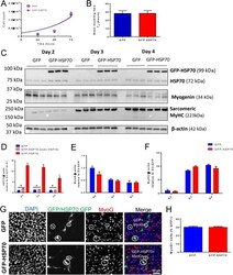
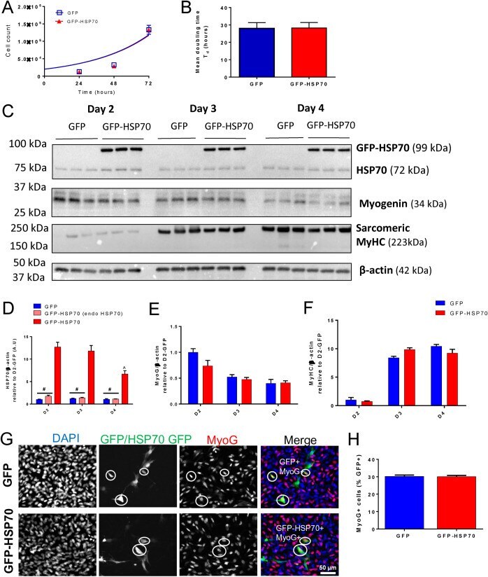
- Fig. 2. GFP-HSP70 overexpression does not alter the rate of C2C12 cell proliferation or differentiation. Plasmid DNA encoding either GFP or a GFP-HSP70 fusion protein were transfected into proliferating C2C12 cells and the effect on proliferation and differentiation was assessed. (A) Total cell counts at 24 h, 48 h and 72 h post-transfection were used to generate exponential growth curves for the GFP and GFP-HSP70 groups. (B) Mean doubling time (T d ) was calculated from the exponential growth curves. Data are presented as mean+-s.e.m. for the cell counts and mean+-95% confidence interval for the doubling time; n =6 replicates/group/timepoint. (C) Representative western blots and quantification of HSP70 (D), myogenin (E), MyHC (F), relative to actin expression in C2C12 cells after 2, 3 and 4 days of differentiation. Data are presented as mean+-s.e.m. and compared with a two-way ANOVA and Tukey's post-hoc test; n =3 replicates/group; # P< 0.05 versus GFP-HSP70 group; ^ P< 0.05 versus D2 GFP-HSP70. (G) Representative immunofluorescence images of C2C12 cells transfected with GFP (top row) or GFP-HSP70 (bottom row) and stained with MyoG at D1. GFP+ MyoG+ cells are circled. (H) The proportion of GFP+ or GFP-HSP70+ cells stained for MyoG was determined. Data are presented as mean+-s.e.m. ; n =3 replicates/group.
- Submitted by
- Invitrogen Antibodies (provider)
- Main image
- Experimental details
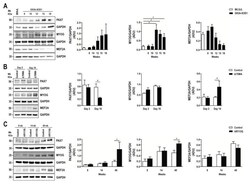
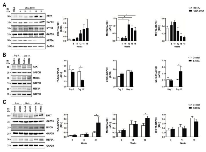
- Figure 3 Altered expression of PAX7, MYOG, and MEF2A proteins in G93A-SOD1, Delta7SMA, and AR113Q mouse muscle as disease progresses. Representative western blot analysis of PAX7, MYOG, and MEF2A proteins in gastrocnemius muscle tissue of ( A ) G93A-SOD1 (black bars), ( B ) Delta7SMA (black bars), and ( C ) AR113Q (black bars) mice and control mice (white bars) ( n = 3 mice per group) with relative densitometric analysis. Density values are reported as mean +- SEM, corrected for background and normalized to GAPDH control. * p < 0.05, Mann-Whitney test.
- Submitted by
- Invitrogen Antibodies (provider)
- Main image
- Experimental details
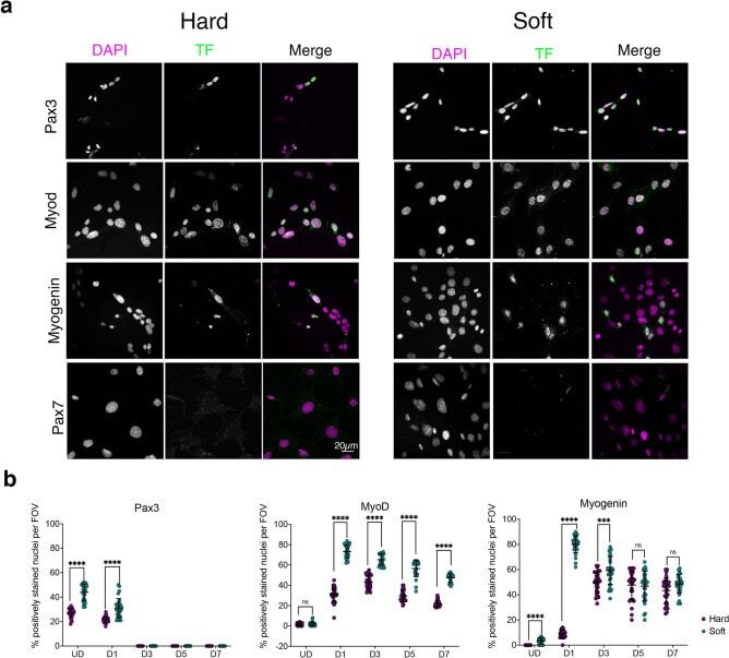
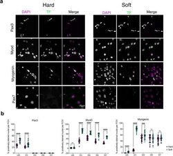
- Figure 4 Immunostaining for myogenic factors in C1F cells cultured on hard or soft surfaces. ( a ) Example images of cells stained for DAPI and specific transcription factor, shown in greyscale for each channel, and as magenta and green respectively in the merged image. ( b ) Quantification of the number of nuclei staining positively for each transcription factor (Pax3, MyoD and myogenin) over time on hard (magenta) and soft (green) surfaces. ****p < 0.0001, ***p < 0.001. ns non-significant.