Explore
 Validate
 Learn
Learn Immunohistochemistry
Antibody data
- Antibody Data
- Antigen structure
- References [1]
- Comments [0]
- Validations
- Immunohistochemistry [1]
- Flow cytometry [1]
- Other assay [3]
Submit
Validation data
Reference
Comment
Report error
- Product number
- MA5-11658 - Provider product page

- Provider
- Invitrogen Antibodies
- Product name
- Myogenin Monoclonal Antibody (F12B)
- Antibody type
- Monoclonal
- Antigen
- Recombinant full-length protein
- Description
- MA5-11658 targets Myogenin in GS, IF, IHC (P), IP, and WB applications and shows reactivity with Human, mouse, and Rat samples.
- Antibody clone number
- F12B
- Concentration
- 0.2 mg/mL
Submitted references Role of Pannexin 1 ATP-Permeable Channels in the Regulation of Signaling Pathways during Skeletal Muscle Unloading.
Zaripova KA, Kalashnikova EP, Belova SP, Kostrominova TY, Shenkman BS, Nemirovskaya TL
International journal of molecular sciences 2021 Sep 28;22(19)
International journal of molecular sciences 2021 Sep 28;22(19)
No comments: Submit comment
Supportive validation
- Submitted by
- Invitrogen Antibodies (provider)
- Main image
- Experimental details
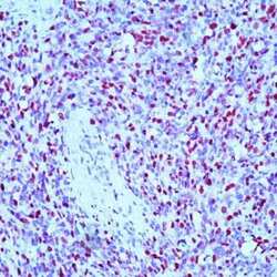
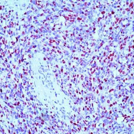
- Formalin-fixed, paraffin-embedded human rhabdomyosarcoma stained with Myogenin antibody using peroxidase-conjugate and AEC chromogen. Note nuclear staining of tumor cells.
Supportive validation
- Submitted by
- Invitrogen Antibodies (provider)
- Main image
- Experimental details
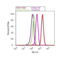
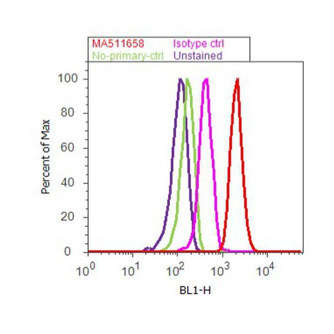
- Flow cytometry analysis of Myogenin was done on C2C12 cells. Cells were fixed with 70% ethanol for 10 minutes, permeabilized with 0.25% Triton™ X-100 for 20 minutes, and blocked with 5% BSA for 30 minutes at room temperature. Cells were labeled with Myogenin Mouse Monoclonal Antibody (MA511658, red histogram) or with mouse isotype control (pink histogram) at 3-5 ug/million cells in 2.5% BSA. After incubation at room temperature for 2 hours, the cells were labeled with Alexa Fluor® 488 Rabbit Anti-Mouse Secondary Antibody (A11059) at a dilution of 1:400 for 30 minutes at room temperature. The representative 10,000 cells were acquired and analyzed for each sample using an Attune® Acoustic Focusing Cytometer. The purple histogram represents unstained control cells and the green histogram represents no-primary-antibody control.
Supportive validation
- Submitted by
- Invitrogen Antibodies (provider)
- Main image
- Experimental details
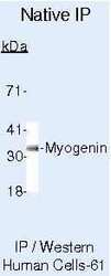
- Immunoprecipitation of Myogenin using Myogenin Monoclonal Antibody (Product # MA5-11658) on Native Human Rh30 Cells.
- Submitted by
- Invitrogen Antibodies (provider)
- Main image

- Experimental details
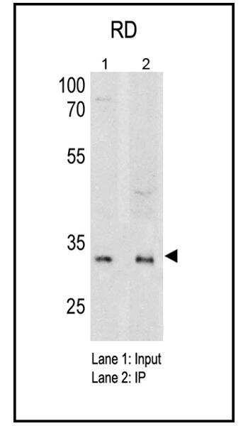
- Immunoprecipitation and Western blot of Myogenin was performed using RD cell lysates. Cells were lysed using ice cold IP lysis/wash buffer and pre-cleared using 50 mL of bead slurry per mL of cell lysate. Antigen-antibody complexes were formed by incubating 0.5 mL pre-cleared cell lysate on ice for 3hrs with 8-15 µg of Myogenin monoclonal antibody (Product # MA5-11658) crosslinked to Protein A/G plus agarose. The immune complexes were eluted using 60 µL sample buffer boiled at 95§C for 5 min and loaded onto an SDS-PAGE gel; Input (lane 1) and Jurkat IP (Lane 2). The membrane was probed with a Myogenin monoclonal antibody (Product # MA5-11658) at a dilution of 3 µg/mg of lysate followed by detection using an HRP-conjugated goat anti-mouse IgG + IgM (H+L) cross-adsorbed secondary antibody. Chemiluminescent detection was performed using an exposure time of 10m, resulting in a ~33 kDa band on input and IP lysates.
- Submitted by
- Invitrogen Antibodies (provider)
- Main image

- Experimental details
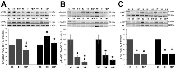
- Figure 3 Evaluation of the MYOG ( A ), p-FoxO3 ( B ) and p-AKT ( C ) content in soleus muscles of non-treated control rats (1C and 3C), rats after 1 and 3 days of unloading (1HS and 3HS), and 1 and 3 days of HS with PRB inhibitor (1HP and 3HP). Values are normalized to the level of GAPDH ( A ), total FoxO3 ( B ) and total Akt ( C ) in each sample. n = 8. * indicates a significant difference from the control, # indicates a significant difference from the HS group, p < 0.05.