Explore
 Validate
 Learn
Learn Western blot
Antibody data
- Antibody Data
- Antigen structure
- References [0]
- Comments [0]
- Validations
- Western blot [1]
- Immunocytochemistry [1]
- Immunohistochemistry [2]
Submit
Validation data
Reference
Comment
Report error
- Product number
- TA328694 - Provider product page

- Provider
- OriGene
- Product name
- Rabbit Polyclonal Anti-A1 Adenosine Receptor
- Antibody type
- Polyclonal
- Description
- Rabbit Polyclonal Anti-A1 Adenosine Receptor
- Host
- Rabbit
- Conjugate
- Unconjugated
- Epitope
- ADORA1
- Antibody clone number
- NULL
- Vial size
- 200 µl
- Concentration
- NULL
No comments: Submit comment
Supportive validation
- Submitted by
- OriGene (provider)
- Main image

- Experimental details
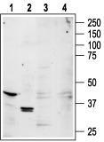
- Western blot analysis of rat brain (lanes 1,3) and rat kidney (lanes 2,4) lysates: 1,2. Anti-A1 Adenosine Receptor antibody, (1:200). 3,4. Anti-A1 Adenosine Receptor antibody, preincubated with the control peptide antigen.
- Validation comment
- WB
Supportive validation
- Submitted by
- OriGene (provider)
- Main image

- Experimental details
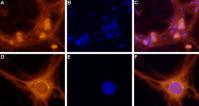
- Expression of A1 Adenosine Receptor in rat DRG primary culture. Immunocytochemical staining of paraformaldehyde-fixed and permeabilized rat dorsal root ganglion (DRG) primary culture. A. Staining of DRG cells with Anti-A1 Adenosine Receptor antibody, (1:100), followed by goat anti-rabbit AlexaFluor-555 secondary antibody. B. Nuclear staining of cells using the cell-permeable dye Hoechst 33342. C. Merged images of A and B. F. Merged images of D and E. Magnification: A-C: x20 D-F: x100
- Validation comment
- IF
Supportive validation
- Submitted by
- OriGene (provider)
- Main image

- Experimental details
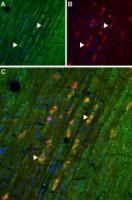
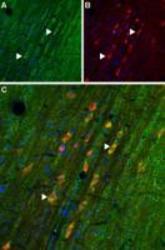
- Expression of A1 Adenosine Receptor in rat cortex Immunohistochemical staining of rat cortex frozen section using Anti-A1 Adenosine Receptor antibody. A. A1 Adenosine Receptor (green) appears in neurons (triangles) and in astrocytes (arrows). B. Parvalbumin staining (red) appears in cortical interneurons. C. Confocal merge of images demonstrates the existence of A1 Adenosine Receptor in a subset of cortical interneurons and astrocytes.
- Validation comment
- IHC
- Submitted by
- OriGene (provider)
- Main image
- Experimental details
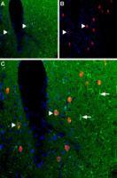
- Expression of A1 Adenosine Receptor in rat medial septum. Immunohistochemical staining of rat medial septum frozen section using Anti-A1 Adenosine Receptor antibody. A. A1 Adenosine Receptor (green) appears in neurons (right pointing triangles). B. Parvalbumin staining (red) appears in medial septal neurons. C. Confocal merge of demonstrates expression of A1 Adenosine Receptor in a subset of medial septal neurons.
- Validation comment
- IHC