Explore
 Validate
 Learn
Learn Western blot
 Immunocytochemistry
Antibody data
- Antibody Data
- Antigen structure
- References [6]
- Comments [0]
- Validations
- Western blot [1]
- Immunohistochemistry [1]
- Flow cytometry [1]
Submit
Validation data
Reference
Comment
Report error
- Product number
- NB300-549 - Provider product page

- Provider
- Novus Biologicals
- Proper citation
- Novus Cat#NB300-549, RRID:AB_10002337
- Product name
- Rabbit Polyclonal Adenosine A1R Antibody
- Antibody type
- Polyclonal
- Description
- Immunogen affinity purified. Detects Adenosine Receptor A1. This does not detect other AR subtypes.
- Reactivity
- Human, Mouse, Rat
- Host
- Rabbit
- Isotype
- IgG
- Vial size
- 100 uL
- Concentration
- 1 mg/ml
- Storage
- Store at -20C. Avoid freeze-thaw cycles.
Submitted references Inhibition of LPS-stimulated ecto-adenosine deaminase attenuates endothelial cell activation.
Adenosine A(1) Receptor mRNA Expression by Neurons and Glia in the Auditory Forebrain.
CD73 is associated with poor prognosis in HNSCC.
Effect of adenosine A1 receptor agonist on the enhanced excitability of spinal dorsal horn neurons after peripheral nerve injury.
Adenosine receptor expression and function in bladder uroepithelium.
Adenosine A(1) receptor-mediated presynaptic inhibition at the calyx of Held of immature rats.
Kutryb-Zajac B, Mierzejewska P, Sucajtys-Szulc E, Bulinska A, Zabielska MA, Jablonska P, Serocki M, Koszalka P, Milczarek R, Jasztal A, Bartoszewski R, Chlopicki S, Slominska EM, Smolenski RT
Journal of molecular and cellular cardiology 2019 Mar;128:62-76
Journal of molecular and cellular cardiology 2019 Mar;128:62-76
Adenosine A(1) Receptor mRNA Expression by Neurons and Glia in the Auditory Forebrain.
Hackett TA
Anatomical record (Hoboken, N.J. : 2007) 2018 Nov;301(11):1882-1905
Anatomical record (Hoboken, N.J. : 2007) 2018 Nov;301(11):1882-1905
CD73 is associated with poor prognosis in HNSCC.
Ren ZH, Lin CZ, Cao W, Yang R, Lu W, Liu ZQ, Chen YM, Yang X, Tian Z, Wang LZ, Li J, Wang X, Chen WT, Ji T, Zhang CP
Oncotarget 2016 Sep 20;7(38):61690-61702
Oncotarget 2016 Sep 20;7(38):61690-61702
Effect of adenosine A1 receptor agonist on the enhanced excitability of spinal dorsal horn neurons after peripheral nerve injury.
Yamaguchi D, Terayama R, Omura S, Tsuchiya H, Sato T, Ichikawa H, Sugimoto T
The International journal of neuroscience 2014 Mar;124(3):213-22
The International journal of neuroscience 2014 Mar;124(3):213-22
Adenosine receptor expression and function in bladder uroepithelium.
Yu W, Zacharia LC, Jackson EK, Apodaca G
American journal of physiology. Cell physiology 2006 Aug;291(2):C254-65
American journal of physiology. Cell physiology 2006 Aug;291(2):C254-65
Adenosine A(1) receptor-mediated presynaptic inhibition at the calyx of Held of immature rats.
Kimura M, Saitoh N, Takahashi T
The Journal of physiology 2003 Dec 1;553(Pt 2):415-26
The Journal of physiology 2003 Dec 1;553(Pt 2):415-26
No comments: Submit comment
Supportive validation
- Submitted by
- Novus Biologicals (provider)
- Main image
- Experimental details
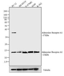
- Western Blot: Adenosine A1R Antibody [NB300-549] - Analysis was performed on whole cell extracts (30 ug lysate) of PC 12 (Lane 1), SH-SY5Y (lane 2), U-87MG (lane 3), A549 (Lane 4) and K-562 (lane 5).

Supportive validation
- Submitted by
- Novus Biologicals (provider)
- Main image
- Experimental details
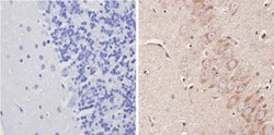
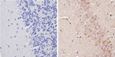
- Immunohistochemistry-Paraffin: Adenosine A1R Antibody [NB300-549] - Analysis showing positive staining in the cytoplasm of Rat brain tissue (right) compared with a negative control in the absence of primary antibody (left).
Supportive validation
- Submitted by
- Novus Biologicals (provider)
- Main image
- Experimental details
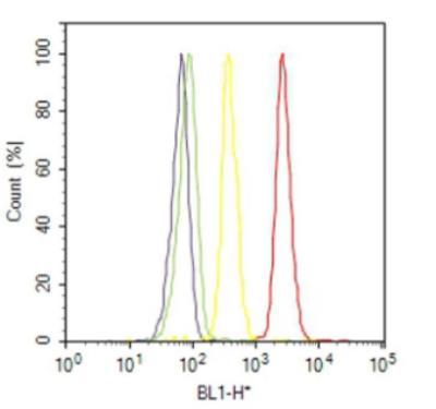
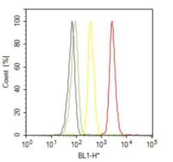
- Flow Cytometry: Adenosine A1R Antibody [NB300-549] - Analysis of Adenosine Receptor A1 was done on A549 cells. Cells were fixed with 70% ethanol for 10 minutes, permeabilized with 0.25% Triton (R) X-100 for 20 minutes, and blocked with 5% BSA for 30 minutes at room temperature. Cells were labeled with Adenosine Receptor A1 Rabbit Polyclonal Antibody (PA1041A, red histogram) or with rabbit isotype control (yellow histogram) at 3-5 ug/million cells in 2.5% BSA. After incubation at room temperature for 2 hours, the cells were labeled with Alexa Fluor (R) 488 Goat Anti-Rabbit Secondary Antibody (A11008) at a dilution of 1:400 for 30 minutes at room temperature. The representative 10,000 cells were acquired and analyzed for each sample using an Attune (R) Acoustic Focusing Cytometer. The purple histogram represents unstained control cells and the green histogram represents no-primary-antibody control.