Explore
 Validate
 Learn
Learn Western blot
 ELISA
ELISAAntibody data
- Antibody Data
- Antigen structure
- References [2]
- Comments [0]
- Validations
- Western blot [2]
- Immunohistochemistry [1]
Submit
Validation data
Reference
Comment
Report error
- Product number
- ABIN359470 - Provider product page

- Provider
- antibodies-online
- Product name
- anti-Calcium/calmodulin-Dependent Protein Kinase IG (CAMK1G) (C-Term) antibody
- Antibody type
- Polyclonal
- Antigen
- Rabbits were immunized wth a KLH conjugated synthetic peptide selected from the C- terminal region of human CaMKIG.
- Description
- Protein G column, eluted with high and low pH buffers and neutralized immediately, followed by dialysis against PBS.
- Reactivity
- Human
- Host
- Rabbit
- Epitope
- C-Term
- Vial size
- 0.4 mL
- Concentration
- 0.25 mg/mL
- Storage
- Store the antibody at 2 - 8°C up to one month or (in aliquots) at -20°C for longer.
- Handling
- Avoid repeated freezing and thawing.
Submitted references Molecular cloning and characterization of CLICK-III/CaMKIgamma, a novel membrane-anchored neuronal Ca2+/calmodulin-dependent protein kinase (CaMK).
A preliminary gene map for the Van der Woude syndrome critical region derived from 900 kb of genomic sequence at 1q32-q41.
Takemoto-Kimura S, Terai H, Takamoto M, Ohmae S, Kikumura S, Segi E, Arakawa Y, Furuyashiki T, Narumiya S, Bito H
The Journal of biological chemistry 2003 May 16;278(20):18597-605
The Journal of biological chemistry 2003 May 16;278(20):18597-605
A preliminary gene map for the Van der Woude syndrome critical region derived from 900 kb of genomic sequence at 1q32-q41.
Schutte BC, Bjork BC, Coppage KB, Malik MI, Gregory SG, Scott DJ, Brentzell LM, Watanabe Y, Dixon MJ, Murray JC
Genome research 2000 Jan;10(1):81-94
Genome research 2000 Jan;10(1):81-94
No comments: Submit comment
Supportive validation
- Submitted by
- antibodies-online (provider)
- Main image

- Experimental details
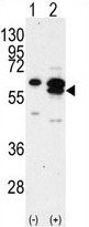
- Western blot analysis of CAMK1G (arrow) using rabbit polyclonal CAMK1G Antibody (C-term) (AP13954PU-N). 293 cell lysates (2 μg/lane) either nontransfected (Lane 1) or transiently transfected with the CAMK1G gene (Lane 2) (Origene Technologies).
- Submitted by
- antibodies-online (provider)
- Main image
- Experimental details
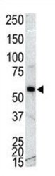
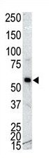
- Western blot analysis of anti-CAMK 1G Pab (AP13954PU-N) in NCI-H460 cell lysate. CAMK 1G (arrow) was detected using purified Pab. Secondary HRP-anti-rabbit was used for signal visualization with chemiluminescence.
Supportive validation
- Submitted by
- antibodies-online (provider)
- Main image

- Experimental details
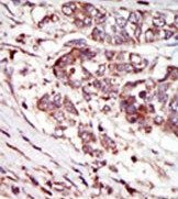
- Formalin-fixed and paraffin-embedded human cancer tissue (hepatocarcinoma) reacted with the primary antibody, which was peroxidase-conjugated to the secondary antibody, followed by AEC staining. This data demonstrates the use of this antibody for immunohistochemistry; clinical relevance has not been evaluated.