Explore
 Validate
 Learn
Learn Western blot
Antibody data
- Antibody Data
- Antigen structure
- References [4]
- Comments [0]
- Validations
- Western blot [1]
- Immunohistochemistry [1]
Submit
Validation data
Reference
Comment
Report error
- Product number
- MAB73461 - Provider product page

- Provider
- Novus Biologicals
- Product name
- Rat Monoclonal TGF-beta 2 Antibody
- Antibody type
- Monoclonal
- Description
- Protein A or G purified from hybridoma culture supernatant. Detects mouse TGF-beta 2 in ELISAs and Western blots. In direct ELISAs, 100% cross-reactivity with recombinant human (rh) TGF-beta 2, 25% cross-reactivity with rhTGF-beta 3, and no cross-reactivity with recombinant mouse TGF-beta 1 is observed.
- Reactivity
- Mouse
- Host
- Rat
- Isotype
- IgG
- Vial size
- 100 ug
- Concentration
- LYOPH
- Storage
- Use a manual defrost freezer and avoid repeated freeze-thaw cycles. 12 months from date of receipt, -20 to -70 degreesC as supplied. 1 month, 2 to 8 degreesC under sterile conditions after reconstitution. 6 months, -20 to -70 degreesC under sterile conditions after reconstitution.
Submitted references Resveratrol protects mice against SEB-induced acute lung injury and mortality by miR-193a modulation that targets TGF-β signalling.
TGF-β1 stimulates movement of renal proximal tubular epithelial cells in a three-dimensional cell culture via an autocrine TGF-β2 production.
Targeting LOXL2 for cardiac interstitial fibrosis and heart failure treatment.
Immunomodulatory cross-talk between conjunctival goblet cells and dendritic cells.
Alghetaa H, Mohammed A, Sultan M, Busbee P, Murphy A, Chatterjee S, Nagarkatti M, Nagarkatti P
Journal of cellular and molecular medicine 2018 May;22(5):2644-2655
Journal of cellular and molecular medicine 2018 May;22(5):2644-2655
TGF-β1 stimulates movement of renal proximal tubular epithelial cells in a three-dimensional cell culture via an autocrine TGF-β2 production.
Luo D, Guan Q, Wang K, Nguan CYC, Du C
Experimental cell research 2017 Jan 1;350(1):132-139
Experimental cell research 2017 Jan 1;350(1):132-139
Targeting LOXL2 for cardiac interstitial fibrosis and heart failure treatment.
Yang J, Savvatis K, Kang JS, Fan P, Zhong H, Schwartz K, Barry V, Mikels-Vigdal A, Karpinski S, Kornyeyev D, Adamkewicz J, Feng X, Zhou Q, Shang C, Kumar P, Phan D, Kasner M, López B, Diez J, Wright KC, Kovacs RL, Chen PS, Quertermous T, Smith V, Yao L, Tschöpe C, Chang CP
Nature communications 2016 Dec 14;7:13710
Nature communications 2016 Dec 14;7:13710
Immunomodulatory cross-talk between conjunctival goblet cells and dendritic cells.
Contreras-Ruiz L, Masli S
PloS one 2015;10(3):e0120284
PloS one 2015;10(3):e0120284
No comments: Submit comment
Supportive validation
- Submitted by
- Novus Biologicals (provider)
- Main image
- Experimental details
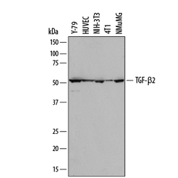
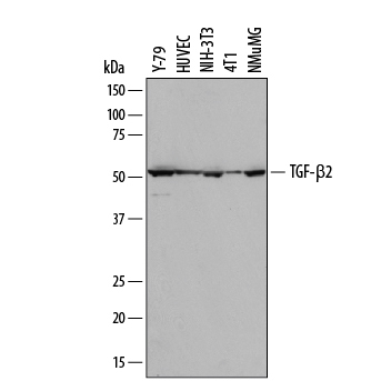
- Detection of Human and Mouse TGF-beta 2 by Western Blot. Western blot shows lysates of Y-79 human retinoblastoma cell line, HUVEC human umbilical vein endothelial cells, NIH-3T3 mouse embryonic fibroblast cell line, 4T1 mouse breast cancer cell line, and NMuMG mouse mammary gland epithelial cell line. PVDF membrane was probed with 2 µg/mL of Rat Anti-Mouse TGF-beta 2 Monoclonal Antibody (Catalog # MAB73461) followed by HRP-conjugated Anti-Rat IgG Secondary Antibody (Catalog # HAF005). A specific band was detected for TGF-beta 2 at approximately 52 kDa (as indicated). This experiment was conducted under reducing conditions and using Immunoblot Buffer Group 1.
Supportive validation
- Submitted by
- Novus Biologicals (provider)
- Main image

- Experimental details
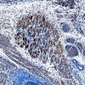
- TGF-beta 2 in Mouse Embryo. TGF-beta 2 was detected in immersion fixed frozen sections of mouse embryo (15 d.p.c.) using Rat Anti-Mouse TGF-beta 2 Monoclonal Antibody (Catalog # MAB73461) at 25 µg/mL overnight at 4 °C. Tissue was stained using the Anti-Rat HRP-DAB Cell & Tissue Staining Kit (brown; Catalog # CTS017) and counterstained with hematoxylin (blue). Specific staining was localized to neuronal processes. View our protocol for Chromogenic IHC Staining of Frozen Tissue Sections.