 Explore
 Validate
 Learn
Learn Western blot
 Flow cytometry
Antibody data
- Antibody Data
- Antigen structure
- References [1]
- Comments [0]
- Validations
- Flow cytometry [2]
- Other assay [1]
Submit
Validation data
Reference
Comment
Report error
- Product number
- 710276 - Provider product page

- Provider
- Invitrogen Antibodies
- Product name
- TGF beta-2 Recombinant Superclonal™ Antibody (12HCLC)
- Antibody type
- Other
- Antigen
- Recombinant full-length protein
- Description
- Recombinant rabbit Superclonal™ antibodies are unique offerings from Thermo Fisher Scientific. They are comprised of a selection of multiple different recombinant monoclonal antibodies, providing the best of both worlds - the sensitivity of polyclonal antibodies with the specificity of monoclonal antibodies - all delivered with the consistency only found in a recombinant antibody. While functionally the same as a polyclonal antibody - recognizing multiple epitope sites on the target and producing higher detection sensitivity for low abundance targets - a recombinant rabbit Superclonal™ antibody has a known mixture of light and heavy chains. The exact population can be produced in every lot, circumventing the biological variability typically associated with polyclonal antibody production. Note: Formerly called “Recombinant polyclonal antibody”, this product is now rebranded as “Recombinant Superclonal™ antibody”. The physical product and the performance remain unchanged.
- Reactivity
- Human, Mouse
- Host
- Rabbit
- Isotype
- IgG
- Antibody clone number
- 12HCLC
- Vial size
- 100 μg
- Concentration
- 0.5 mg/mL
- Storage
- Store at 4°C short term. For long term storage, store at -20°C, avoiding freeze/thaw cycles.
Submitted references Dexamethasone-induced inhibition of miR-132 via methylation promotes TGF-β-driven progression of pancreatic cancer.
Abukiwan A, Nwaeburu CC, Bauer N, Zhao Z, Liu L, Gladkich J, Gross W, Benner A, Strobel O, Fellenberg J, Herr I
International journal of oncology 2019 Jan;54(1):53-64
International journal of oncology 2019 Jan;54(1):53-64
No comments: Submit comment
Supportive validation
- Submitted by
- Invitrogen Antibodies (provider)
- Main image
- Experimental details
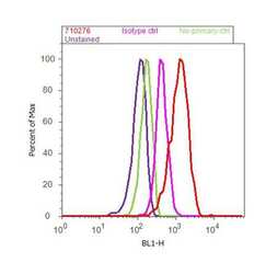
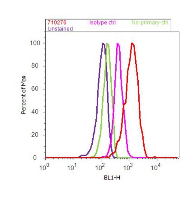
- Flow cytometry analysis of TGF beta-2 was done on U-87 MG cells. Cells were fixed with 70% ethanol for 10 minutes, permeabilized with 0.25% Triton™ X-100 for 20 minutes, and blocked with 5% BSA for 30 minutes at room temperature. Cells were labeled with TGF beta-2 Recombinat Rabbit Polyclonal Antibody (Product # 710276, red histogram) or with rabbit isotype control (pink histogram) at 3-5 µg/million cells in 2.5% BSA. After incubation at room temperature for 2 hours, the cells were labeled with Alexa Fluor® 488 Goat anti-Rabbit Secondary Antibody (Product # A-11008) at a dilution of 1:400 for 30 minutes at room temperature. The representative 10,000 cells were acquired and analyzed for each sample using an Attune® Acoustic Focusing Cytometer. The purple histogram represents unstained control cells and the green histogram represents no-primary-antibody control.

- Submitted by
- Invitrogen Antibodies (provider)
- Main image
- Experimental details
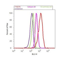
- Flow cytometry analysis of TGF beta-2 was done on U-87 MG cells. Cells were fixed with 70% ethanol for 10 minutes, permeabilized with 0.25% Triton™ X-100 for 20 minutes, and blocked with 5% BSA for 30 minutes at room temperature. Cells were labeled with TGF beta-2 Recombinat Rabbit Polyclonal Antibody (Product # 710276, red histogram) or with rabbit isotype control (pink histogram) at 3-5 µg/million cells in 2.5% BSA. After incubation at room temperature for 2 hours, the cells were labeled with Alexa Fluor® 488 Goat anti-Rabbit Secondary Antibody (Product # A-11008) at a dilution of 1:400 for 30 minutes at room temperature. The representative 10,000 cells were acquired and analyzed for each sample using an Attune® Acoustic Focusing Cytometer. The purple histogram represents unstained control cells and the green histogram represents no-primary-antibody control.
Supportive validation
- Submitted by
- Invitrogen Antibodies (provider)
- Main image
- Experimental details
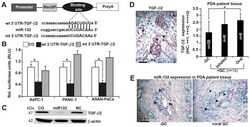
- Figure 3 miR-132 binds to the 3'UTR of TGF-beta2 to inhibit its expression. (A) Schematic of the miR-132 binding site in the TGF-beta2 3'UTR at position 746-753 and its sequence homology to miR-132. The mutant version of the TGF-beta2 3'UTR is shown. (B) The wt and mt TGF-beta2 3'UTRs were cloned into a pLightSwitch Renilla plasmid and transfected into AsPC-1, PANC-1 and ASAN-PaCa cells in the presence or absence of 50 nM miR-132 mimics. Negative mimics served as a control. Co-transfection with Firefly luciferase (0.25 ng/ u l) served as a normalization control. At 48 h after transfection, the expression of Renilla and Firefly luciferases was detected using a FLUOstar Omega microplate reader. Renilla luciferase activities were normalized to Firefly luciferase activities. (C) PANC-1 cells were transfected with miR-132 or non-coding miRNA (NC), or mock-treated without miRNA (CO). Proteins were harvested 48 h later and analyzed by western blotting. beta-actin served as the normalization control. (D) Representative paraffin-embedded PDA tissue sections from patients with documented pre-operative intake of inhaled (n=8) or oral (n=6) GCs (+GC, n=14) and from those without GC intake (-GC, n=20) were evaluated by IHC to detect the expression of TGF-beta2. A semi-quantitative scoring system was used to evaluate expression levels based on visual determination of the percentage of positive cells. The sections were analyzed at x400 magnification, and representative images are s
