Explore
 Validate
 Learn
Learn Other assay
Antibody data
- Antibody Data
- Antigen structure
- References [3]
- Comments [0]
- Validations
- Other assay [1]
Submit
Validation data
Reference
Comment
Report error
- Product number
- PAB0517 - Provider product page

- Provider
- Abnova Corporation
- Proper citation
- Abnova Corporation Cat#PAB0517, RRID:AB_982696
- Product name
- MAP3K7 (phospho T187) polyclonal antibody
- Antibody type
- Polyclonal
- Description
- Rabbit polyclonal antibody raised against synthetic phosphopeptide of MAP3K7.
- Storage
- Store at 4°C. For long term storage store at -20°C.Aliquot to avoid repeated freezing and thawing.
Submitted references Wnt activates the Tak1/Nemo-like kinase pathway.
Tumor necrosis factor-alpha-induced IKK phosphorylation of NF-kappaB p65 on serine 536 is mediated through the TRAF2, TRAF5, and TAK1 signaling pathway.
A dominant negative TAK1 inhibits cellular fibrotic responses induced by TGF-beta.
Smit L, Baas A, Kuipers J, Korswagen H, van de Wetering M, Clevers H
The Journal of biological chemistry 2004 Apr 23;279(17):17232-40
The Journal of biological chemistry 2004 Apr 23;279(17):17232-40
Tumor necrosis factor-alpha-induced IKK phosphorylation of NF-kappaB p65 on serine 536 is mediated through the TRAF2, TRAF5, and TAK1 signaling pathway.
Sakurai H, Suzuki S, Kawasaki N, Nakano H, Okazaki T, Chino A, Doi T, Saiki I
The Journal of biological chemistry 2003 Sep 19;278(38):36916-23
The Journal of biological chemistry 2003 Sep 19;278(38):36916-23
A dominant negative TAK1 inhibits cellular fibrotic responses induced by TGF-beta.
Ono K, Ohtomo T, Ninomiya-Tsuji J, Tsuchiya M
Biochemical and biophysical research communications 2003 Jul 25;307(2):332-7
Biochemical and biophysical research communications 2003 Jul 25;307(2):332-7
No comments: Submit comment
Supportive validation
- Submitted by
- Abnova Corporation (provider)
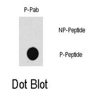
- Main image

- Experimental details
- Dot blot analysis of MAP3K7 (phospho T187) polyclonal antibody (Cat # PAB0517) on nitrocellulose membrane. 50 ng of Phospho-peptide or Non Phospho-peptide per dot were adsorbed. Antibody working concentration were 0.5 ug/mL.
- Validation comment
- Dot Blot (Peptide)