 Explore
 Validate
 Learn
Learn Western blot
 Flow cytometry
Antibody data
- Antibody Data
- Antigen structure
- References [3]
- Comments [0]
- Validations
- Western blot [3]
Submit
Validation data
Reference
Comment
Report error
- Product number
- AF397 - Provider product page

- Provider
- R&D Systems
- Product name
- Human/Mouse/Rat TrkB Antibody
- Antibody type
- Polyclonal
- Description
- Antigen Affinity-purified. Detects human TrkB in direct ELISA. Detects human, mouse, and rat TrkB Western blots. In direct ELISA and Western blots, less than 1% cross-reactivity with recombinant human (rh) TrkC, rhNT-3, rhNT-4, rhBDNF, and rh beta-NGF is observed.
- Reactivity
- Human, Mouse, Rat
- Host
- Goat
- Conjugate
- Unconjugated
- Isotype
- IgG
- Vial size
- 100 ug
- Concentration
- LYOPH
- Storage
- Use a manual defrost freezer and avoid repeated freeze-thaw cycles. 12 months from date of receipt, -20 to -70 °C as supplied. 1 month, 2 to 8 °C under sterile conditions after reconstitution. 6 months, -20 to -70 °C under sterile conditions after reconstitution.
Submitted references The effects of rotenone on TH, BDNF and BDNF-related proteins in the brain and periphery: Relevance to early Parkinson's disease.
Constitutively active TrkB confers an aggressive transformed phenotype to a neural crest-derived cell line.
Protein tyrosine phosphatase receptor type O inhibits trigeminal axon growth and branching by repressing TrkB and Ret signaling.
Johnson ME, Zhou XF, Bobrovskaya L
Journal of chemical neuroanatomy 2019 Apr;97:23-32
Journal of chemical neuroanatomy 2019 Apr;97:23-32
Constitutively active TrkB confers an aggressive transformed phenotype to a neural crest-derived cell line.
Dewitt J, Ochoa V, Urschitz J, Elston M, Moisyadi S, Nishi R
Oncogene 2014 Feb 20;33(8):977-85
Oncogene 2014 Feb 20;33(8):977-85
Protein tyrosine phosphatase receptor type O inhibits trigeminal axon growth and branching by repressing TrkB and Ret signaling.
Gatto G, Dudanova I, Suetterlin P, Davies AM, Drescher U, Bixby JL, Klein R
The Journal of neuroscience : the official journal of the Society for Neuroscience 2013 Mar 20;33(12):5399-410
The Journal of neuroscience : the official journal of the Society for Neuroscience 2013 Mar 20;33(12):5399-410
No comments: Submit comment
Supportive validation
- Submitted by
- R&D Systems (provider)
- Main image

- Experimental details
- Detection of Human TrkB by Western Blot. Western blot shows lysates of human brain (motor cortex) tissue, human brain (cerebellum) tissue, and human brain (hypothalamus) tissue. PVDF membrane was probed with 0.5 µg/mL of Goat Anti-Human/Mouse/Rat TrkB Antigen Affinity-purified Polyclonal Antibody (Catalog # AF397) followed by HRP-conjugated Anti-Goat IgG Secondary Antibody (Catalog # HAF017). Specific bands were detected for TrkB at approximately 90-100 & 140 kDa (as indicated). This experiment was conducted under reducing conditions and using Immunoblot Buffer Group 1.

- Submitted by
- R&D Systems (provider)
- Main image

- Experimental details
- Detection of Human TrkB by Simple WesternTM. Simple Western lane view shows lysates of human brain (cerebellum) tissue, loaded at 0.2 mg/mL. A specific band was detected for TrkB at approximately 162 kDa (as indicated) using 10 µg/mL of Goat Anti-Human/Mouse/Rat TrkB Antigen Affinity-purified Polyclonal Antibody (Catalog # AF397) followed by 1:50 dilution of HRP-conjugated Anti-Goat IgG Secondary Antibody (Catalog # HAF109). This experiment was conducted under reducing conditions and using the 12-230 kDa separation system.

- Submitted by
- R&D Systems (provider)
- Main image
- Experimental details
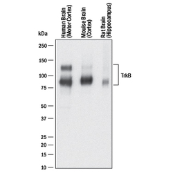
- Detection of Human, Mouse, and Rat TrkB by Western Blot. Western blot shows lysates of human brain (motor cortex) tissue, mouse brain (cortex) tissue, and rat brain (hippocampus) tissue. PVDF membrane was probed with 0.25 µg/mL of Goat Anti-Human/Mouse/Rat TrkB Antigen Affinity-purified Polyclonal Antibody (Catalog # AF397) followed by HRP-conjugated Anti-Goat IgG Secondary Antibody (Catalog # HAF017). Specific bands were detected for TrkB at approximately 95 kDa and 145 kDa (as indicated). This experiment was conducted under reducing conditions and using Immunoblot Buffer Group 1.
