Explore
 Validate
 Learn
Learn Western blot
Antibody data
- Antibody Data
- Antigen structure
- References [3]
- Comments [0]
- Validations
- Western blot [1]
- Immunohistochemistry [1]
Submit
Validation data
Reference
Comment
Report error
- Product number
- PAB9938 - Provider product page

- Provider
- Abnova Corporation
- Proper citation
- Abnova Corporation Cat#PAB9938, RRID:AB_1672184
- Product name
- CAMK4 polyclonal antibody
- Antibody type
- Polyclonal
- Description
- Rabbit polyclonal antibody raised against synthetic peptide of CAMK4.
- Storage
- Store at 4°C on dry atmosphere.After reconstitution with 0.1 mL of deionized water, store at -20°C or lower.Aliquot to avoid repeated freezing and thawing.
Submitted references The cDNA sequence and characterization of the Ca2+/calmodulin-dependent protein kinase-Gr from human brain and thymus.
A Ca2+/calmodulin-dependent protein kinase, CaM kinase-Gr, expressed after transformation of primary human B lymphocytes by Epstein-Barr virus (EBV) is induced by the EBV oncogene LMP1.
cDNA cloning and expression of human calmodulin-dependent protein kinase IV.
Bland MM, Monroe RS, Ohmstede CA
Gene 1994 May 16;142(2):191-7
Gene 1994 May 16;142(2):191-7
A Ca2+/calmodulin-dependent protein kinase, CaM kinase-Gr, expressed after transformation of primary human B lymphocytes by Epstein-Barr virus (EBV) is induced by the EBV oncogene LMP1.
Mosialos G, Hanissian SH, Jawahar S, Vara L, Kieff E, Chatila TA
Journal of virology 1994 Mar;68(3):1697-705
Journal of virology 1994 Mar;68(3):1697-705
cDNA cloning and expression of human calmodulin-dependent protein kinase IV.
Kitani T, Okuno S, Fujisawa H
Journal of biochemistry 1994 Apr;115(4):637-40
Journal of biochemistry 1994 Apr;115(4):637-40
No comments: Submit comment
Supportive validation
- Submitted by
- Abnova Corporation (provider)
- Main image
- Experimental details
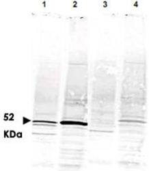
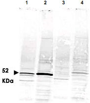
- Western blot using CAMK4 polyclonal antibody (Cat # PAB9938) shows detection of a band ~52 KDa corresponding to CAMK4 (arrowhead) in various preparations.Staining ofrat brain lysate is shown in lane 1.Jurkat cell lysate staining is shown in lane 2.Specific reactivity is blocked in both lysates when antibodyis preincubated with immunizing peptide (lanes 3 and 4 respectively).Approximately 35 ug of each lysate was separated by 4-20% SDS-PAGE and transferred onto nitrocellulose.CAMK4 was similarly detected on lysates from mouse brain (not shown).After blocking the membrane was probed with the primary antibody diluted to 1 : 1,000 for 2h at room temperature followed by washes and reaction with a 1 : 10,000 dilution of IRDye™800 conjugated Gt-a-Rabbit IgG [H&L]MX for 45 min at room temperature.
Supportive validation
- Submitted by
- Abnova Corporation (provider)
- Main image
- Experimental details
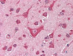
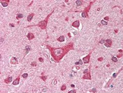
- Immunohistochemical staining with CAMK4 polyclonal antibody (Cat # PAB9938) was diluted 1 : 500 to detect CAMK4 in human brain cortex tissue.Tissue was formalin fixed and paraffin embedded.No pre-treatment of sample was required. The image shows the localization of antibody as the precipitated red signal, with a hematoxylin purple nuclear counter stain.
- Validation comment
- Immunohistochemistry (Formalin/PFA-fixed paraffin-embedded sections)