Explore
 Validate
 Learn
Learn Western blot
 ELISA
ELISAAntibody data
- Antibody Data
- Antigen structure
- References [0]
- Comments [0]
- Validations
- Western blot [1]
- Immunohistochemistry [1]
Submit
Validation data
Reference
Comment
Report error
- Product number
- NBP1-78002 - Provider product page

- Provider
- Novus Biologicals
- Proper citation
- Novus Cat#NBP1-78002, RRID:AB_11047801
- Product name
- Rabbit Polyclonal CaMKIV Antibody
- Antibody type
- Polyclonal
- Description
- Delipidation and Defibrination. This antiserum is directed against human CaM Kinase IV protein.A BLAST analysis was used to suggest reactivity with this protein.
- Reactivity
- Human, Mouse, Rat, Bovine, Canine
- Host
- Rabbit
- Vial size
- 0.1 ml
- Concentration
- LYOPH
- Storage
- Store lyophilized antibody at 4C. Aliquot reconstituted liquid and store at -20C. Avoid freeze-thaw cycles.
No comments: Submit comment
Supportive validation
- Submitted by
- Novus Biologicals (provider)
- Main image
- Experimental details
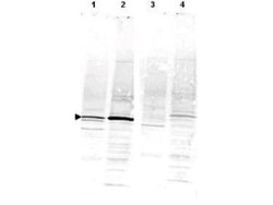
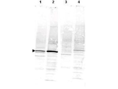
- Western Blot: CaMKIV Antibody [NBP1-78002] - Shows detection of a band ~52 kDa corresponding to CaM Kinase IV (arrowhead) in various preparations: lane 1 - rat brain lysate, lane 2 - Jurkat cell lysate. Specific reactivity is blocked in both lysates when antibody is preincubated with immunizing peptide (lanes 3 and 4 respectively). Approximately 35 ug of each lysate was separated by 4-20% SDS-PAGE and transferred onto nitrocellulose. CaM Kinase IV was similarly detected on lysates from mouse brain (not shown). After blocking the membrane was probed with the primary antibody diluted to 1:1,000 for 2h at room temperature followed by washes and reaction with a 1:10,000 dilution of IRDye800 conjugated Gt-a-Rabbit IgG [H&L] MX for 45 min at room temperature.
Supportive validation
- Submitted by
- Novus Biologicals (provider)
- Main image
- Experimental details
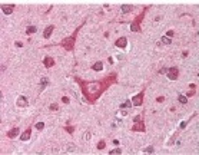
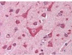
- Immunohistochemistry-Paraffin: CaMKIV Antibody [NBP1-78002] - Antibody was diluted 1:500 in human brain cortex tissue. Tissue was formalin fixed and paraffin embedded. No pre-treatment of sample was required. The image shows the localization of antibody as the precipitated red signal, with a hematoxylin purple nuclear counter stain.