Explore
 Validate
 Learn
Learn Western blot
 ELISA
ELISAAntibody data
- Antibody Data
- Antigen structure
- References [0]
- Comments [0]
- Validations
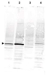
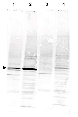
- Western blot [1]
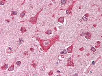
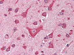
- Immunohistochemistry [1]
Submit
Validation data
Reference
Comment
Report error
- Product number
- AP09079SU-N - Provider product page

- Provider
- Acris Antibodies GmbH
- Proper citation
- Acris Antibodies GmbH Cat#AP09079SU-N, RRID:AB_2035240
- Product name
- anti CAMK4 (305-323)
- Antibody type
- Polyclonal
- Antigen
- Synthetic peptide corresponding to amino acids 305-323 of Human CaM Kinase IV protein
- Reactivity
- Human, Mouse, Rat, Bovine, Canine, Chicken/Avian, Zebrafish
- Host
- Rabbit
- Isotype
- IgG
- Vial size
- 0.1 ml
- Concentration
- 70 mg/ml by Refractometry
No comments: Submit comment
Supportive validation
- Submitted by
- Acris Antibodies GmbH (provider)
- Main image
- Experimental details
- Western blot using Anti-CaM Kinase IV antibody shows detection of a band ~52 kDa corresponding to CaM Kinase IV (arrowhead) in various preparations. Staining of rat brain lysate is shown in lane 1. Jurkat cell lysate staining is shown in lane 2. Specific reactivity is blocked in both lysates when antibody is preincubated with immunizing peptide (lanes 3 and 4 respectively). Approximately 35 µg of each lysate was separated by 4-20% SDS-PAGE and transferred onto nitrocellulose. CaM Kinase IV was similarly detected on lysates from mouse brain (not shown). After blocking the membrane was probed with the primary antibody diluted to 1:1,000 for 2h at room temperature followed by washes and reaction with a 1:10,000 dilution of IRDye(TM)800 conjugated Gt-a-Rabbit IgG [H&L] MX for 45 min at room temperature. IRDye(TM)800 fluorescence image was captured using the Odyssey(R) Infrared Imaging System developed by LI-COR.
Supportive validation
- Submitted by
- Acris Antibodies GmbH (provider)
- Main image
- Experimental details
- Immunohistochemistry. Anti-CAMK4 antibody was diluted 1:500 to detect CAMK4 in human brain cortex tissue. Tissue was formalin fixed and paraffin embedded. No pre-treatment of sample was required. The image shows the localization of antibody as the precipitated red signal, with a hematoxylin purple nuclear counter stain.