 Explore
 Validate
 Learn
Learn Western blot
 Immunohistochemistry
Antibody data
- Antibody Data
- Antigen structure
- References [2]
- Comments [0]
- Validations
- Immunohistochemistry [1]
- Other assay [2]
Submit
Validation data
Reference
Comment
Report error
- Product number
- PA5-15181 - Provider product page

- Provider
- Invitrogen Antibodies
- Product name
- TNIK Polyclonal Antibody
- Antibody type
- Polyclonal
- Antigen
- Synthetic peptide
- Reactivity
- Human, Rat
- Host
- Rabbit
- Isotype
- IgG
- Vial size
- 400 μL
- Concentration
- 2 mg/mL
- Storage
- Store at 4°C short term. For long term storage, store at -20°C, avoiding freeze/thaw cycles.
Submitted references A null mutation in TNIK defines a novel locus for intellectual disability.
Organization of TNIK in dendritic spines.
Anazi S, Shamseldin HE, AlNaqeb D, Abouelhoda M, Monies D, Salih MA, Al-Rubeaan K, Alkuraya FS
Human genetics 2016 Jul;135(7):773-8
Human genetics 2016 Jul;135(7):773-8
Organization of TNIK in dendritic spines.
Burette AC, Phend KD, Burette S, Lin Q, Liang M, Foltz G, Taylor N, Wang Q, Brandon NJ, Bates B, Ehlers MD, Weinberg RJ
The Journal of comparative neurology 2015 Sep 1;523(13):1913-24
The Journal of comparative neurology 2015 Sep 1;523(13):1913-24
No comments: Submit comment
Supportive validation
- Submitted by
- Invitrogen Antibodies (provider)
- Main image

- Experimental details
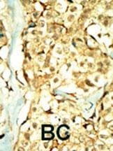
- Immunohistochemistry analysis of TNIK in formalin-fixed and paraffin-embedded human cancer tissue. Samples were incubated with TNIK polyclonal antibody (Product # PA5-15181) which was peroxidase-conjugated to the secondary antibody, followed by DAB staining. This data demonstrates the use of this antibody for immunohistochemistry; clinical relevance has not been evaluated. BC = breast carcinoma; HC = hepatocarcinoma.
Supportive validation
- Submitted by
- Invitrogen Antibodies (provider)
- Main image

- Experimental details
- NULL
- Submitted by
- Invitrogen Antibodies (provider)
- Main image
- Experimental details
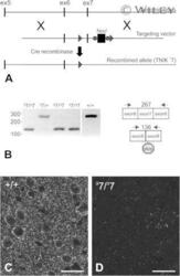
- Preparation of the knockout mice. A: Gene targeting of TNIK locus and RT-PCR of whole brain mRNA from targeted mice. Genomic locus illustrating the region of exons 5-7. Targeting vector indicated showing homology arms, LoxP sites flanking exon 7 (>), and neomycin (G418) resistance cassette used for selection. Neo r cassette is flanked by Frt sites (*). After introduction into the germline, exon 7 was removed by Cre recombinase-mediated deletion. B: RT-PCR of whole brain RNA from animals of the indicated genotypes. PCR primers located in exons 6 and 8 are indicated by arrows. Deletion of exon 7 results in a novel transcript in animals carrying the Delta7 allele that contains early termination codons as indicated. Identity of PCR products was verified by sequencing (data not shown). C,D: Immunofluorescence staining for TNIK in neocortex from WT (C) and TNIK Delta7 /TNIK Delta7 (D) mouse brain. Scale bar = 50 um in C and D.
