Explore
 Validate
 Learn
Learn Immunocytochemistry
 Immunohistochemistry
Antibody data
- Antibody Data
- Antigen structure
- References [3]
- Comments [0]
- Validations
- Immunocytochemistry [1]
Submit
Validation data
Reference
Comment
Report error
- Product number
- HPA012128 - Provider product page

- Provider
- Atlas Antibodies
- Proper citation
- Atlas Antibodies Cat#HPA012128, RRID:AB_1858226
- Product name
- Anti-TNIK
- Antibody type
- Polyclonal
- Description
- Polyclonal Antibody against Human TNIK, Gene description: TRAF2 and NCK interacting kinase, Alternative Gene Names: KIAA0551, Validated applications: ICC, IHC, Uniprot ID: Q9UKE5, Storage: Store at +4°C for short term storage. Long time storage is recommended at -20°C.
- Reactivity
- Human
- Host
- Rabbit
- Conjugate
- Unconjugated
- Isotype
- IgG
- Vial size
- 100 µl
- Concentration
- 0.1 mg/ml
- Storage
- Store at +4°C for short term storage. Long time storage is recommended at -20°C.
- Handling
- The antibody solution should be gently mixed before use.
Submitted references Organization of TNIK in dendritic spines
Prognostic significance of Traf2- and Nck- interacting kinase (TNIK) in colorectal cancer
The essential role of TNIK gene amplification in gastric cancer growth
Burette A, Phend K, Burette S, Lin Q, Liang M, Foltz G, Taylor N, Wang Q, Brandon N, Bates B, Ehlers M, Weinberg R
Journal of Comparative Neurology 2015;523(13):1913-1924
Journal of Comparative Neurology 2015;523(13):1913-1924
Prognostic significance of Traf2- and Nck- interacting kinase (TNIK) in colorectal cancer
Takahashi H, Ishikawa T, Ishiguro M, Okazaki S, Mogushi K, Kobayashi H, Iida S, Mizushima H, Tanaka H, Uetake H, Sugihara K
BMC Cancer 2015;15(1)
BMC Cancer 2015;15(1)
The essential role of TNIK gene amplification in gastric cancer growth
Yu D, Zhang X, Wang H, Zhang L, Chen H, Hu M, Dong Z, Zhu G, Qian Z, Fan J, Su X, Xu Y, Zheng L, Dong H, Yin X, Ji Q, Ji J
Oncogenesis 2014;3(2):e89-e89
Oncogenesis 2014;3(2):e89-e89
No comments: Submit comment
Supportive validation
- Submitted by
- Atlas Antibodies (provider)
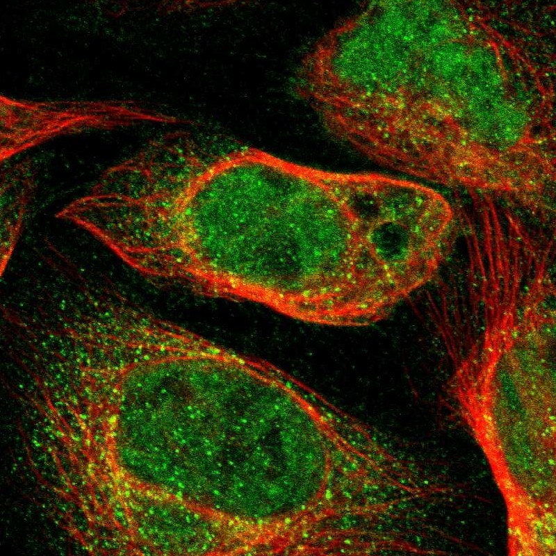
- Main image

- Experimental details
- Immunofluorescent staining of human cell line U-2 OS shows localization to nucleoplasm & cytosol.
- Sample type
- Human