Explore
 Validate
 Learn
Learn Immunocytochemistry
Antibody data
- Antibody Data
- Antigen structure
- References [1]
- Comments [0]
- Validations
- Immunocytochemistry [2]
Submit
Validation data
Reference
Comment
Report error
- Product number
- PA3-851 - Provider product page

- Provider
- Invitrogen Antibodies
- Product name
- Anti-IRAG
- Antibody type
- Polyclonal
- Antigen
- Synthetic peptide corresponding to residues 93-107 of bovine IRAG.
- Host
- Rabbit
- Vial size
- 100 µl
- Storage
- -20° C, Avoid Freeze/Thaw Cycles
Submitted references Regulation of intracellular calcium by a signalling complex of IRAG, IP3 receptor and cGMP kinase Ibeta.
Schlossmann J, Ammendola A, Ashman K, Zong X, Huber A, Neubauer G, Wang GX, Allescher HD, Korth M, Wilm M, Hofmann F, Ruth P
Nature 2000 Mar 9;404(6774):197-201
Nature 2000 Mar 9;404(6774):197-201
No comments: Submit comment
Supportive validation
- Submitted by
- Invitrogen Antibodies (provider)
- Main image
- Experimental details
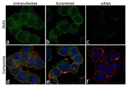
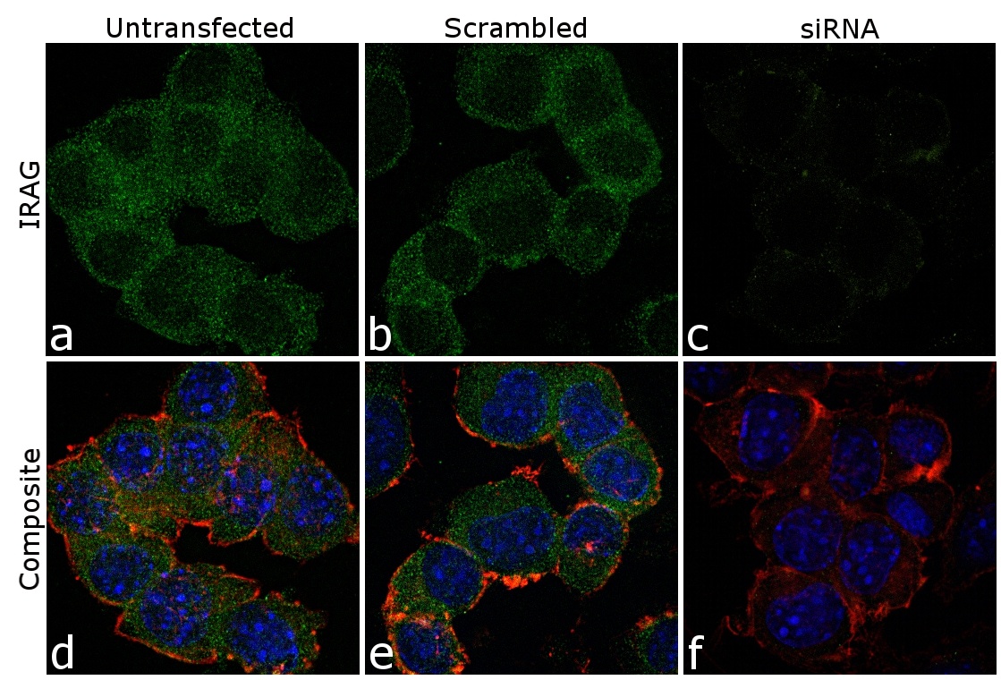
- Knockdown of MRVI1 was achieved by transfecting Neuro-2a cells with MRVI1 specific siRNA (Silencer® select Product # 70042 and s70043). Immunofluorescence analysis was performed on Neuro-2a cells (untransfected, panel a,d), transfected with non-specific scrambled siRNA (panels b,e) and transfected with MRVI1 specific siRNA (panel c,f). Cells were fixed, permeabilized, and labelled with MRVI1 Rabbit Polyclonal Antibody (Product # PA3-851, 1:250 dilution), followed by Goat anti-Rabbit IgG (Heavy Chain) Superclonal™ Secondary Antibody, Alexa Fluor® 488 conjugate (Product # A27034, 1:2000 dilution). Nuclei (blue) were stained using SlowFade® Gold Antifade Mountant with DAPI (Product # S36938), and Rhodamine Phalloidin (Product # R415, 1:300) was used for cytoskeletal F-actin (red) staining. Loss of signal was observed upon siRNA mediated knockdown (panel c,f) confirming specificity of the antibody to MRVI1 (green). The images were captured at 60X magnification.
- Submitted by
- Invitrogen Antibodies (provider)
- Main image
- Experimental details
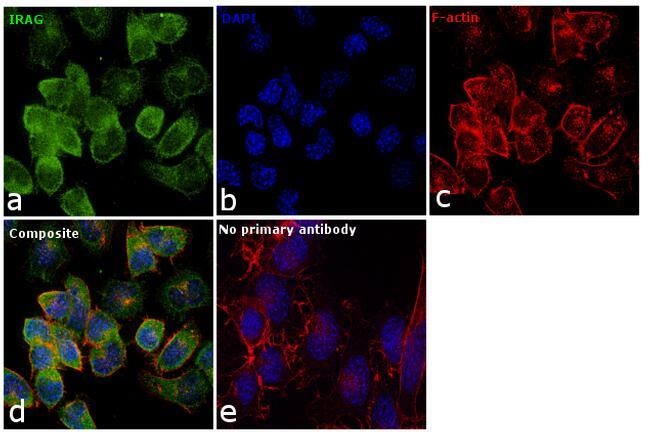
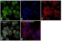
- Immunofluorescence analysis of MRVI1 (IRAG) was performed using 70% confluent log phase Neuro-2a cells. The cells were fixed with 4% paraformaldehyde for 10 minutes, permeabilized with 0.1% Triton™ X-100 for 10 minutes, and blocked with 1% BSA for 1 hour at room temperature. The cells were labeled with MRVI1 Rabbit polyclonal antibody (Product # PA3-851) at 5 µg/mL in 0.1% BSA, incubated overnight at 4 degree Celsius and then labeled with Goat anti-Rabbit IgG (Heavy Chain) Superclonal™ Secondary Antibody, Alexa Fluor® 488 conjugate (Product # A27034) at a dilution of 1:2000 for 45 minutes at room temperature (Panel a: green). Nuclei (Panel b: blue) were stained with SlowFade® Gold Antifade Mountant with DAPI (Product # S36938). F-actin (Panel c: red) was stained with Rhodamine Phalloidin (Product # R415, 1:300). Panel d represents the merged image showing cytoplasmic and membranous localization. Panel e shows control cells with no primary antibody to assess background. The images were captured at 60X magnification.