Explore
 Validate
 Learn
Learn Western blot
Antibody data
- Antibody Data
- Antigen structure
- References [4]
- Comments [0]
- Validations
- Western blot [3]
- Immunohistochemistry [5]
Submit
Validation data
Reference
Comment
Report error
- Product number
- GTX114462 - Provider product page

- Provider
- GeneTex
- Proper citation
- GeneTex Cat#GTX114462, RRID:AB_11173658
- Product name
- Histone H1.0 antibody
- Antibody type
- Polyclonal
- Reactivity
- Human, Mouse, Rat
- Host
- Rabbit
Submitted references Temporally and Spatially Regulated Expression of the Linker Histone H1fx During Mouse Development.
Nucleosome-nucleosome interactions via histone tails and linker DNA regulate nuclear rigidity.
Pigment Epithelium-Derived Factor Mediates Autophagy and Apoptosis in Myocardial Hypoxia/Reoxygenation Injury.
Celastrol prevents circulatory failure via induction of heme oxygenase-1 and heat shock protein 70 in endotoxemic rats.
Ichihara-Tanaka K, Kadomatsu K, Kishida S
The journal of histochemistry and cytochemistry : official journal of the Histochemistry Society 2017 Sep;65(9):513-530
The journal of histochemistry and cytochemistry : official journal of the Histochemistry Society 2017 Sep;65(9):513-530
Nucleosome-nucleosome interactions via histone tails and linker DNA regulate nuclear rigidity.
Shimamoto Y, Tamura S, Masumoto H, Maeshima K
Molecular biology of the cell 2017 Jun 1;28(11):1580-1589
Molecular biology of the cell 2017 Jun 1;28(11):1580-1589
Pigment Epithelium-Derived Factor Mediates Autophagy and Apoptosis in Myocardial Hypoxia/Reoxygenation Injury.
Kuo HF, Liu PL, Chong IW, Liu YP, Chen YH, Ku PM, Li CY, Chen HH, Chiang HC, Wang CL, Chen HJ, Chen YC, Hsieh CC
PloS one 2016;11(5):e0156059
PloS one 2016;11(5):e0156059
Celastrol prevents circulatory failure via induction of heme oxygenase-1 and heat shock protein 70 in endotoxemic rats.
Wang YL, Lam KK, Cheng PY, Lee YM
Journal of ethnopharmacology 2015 Mar 13;162:168-75
Journal of ethnopharmacology 2015 Mar 13;162:168-75
No comments: Submit comment
Supportive validation
- Submitted by
- GeneTex (provider)
- Main image
- Experimental details
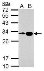
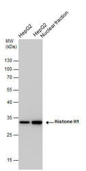
- Sample (30 ug of whole cell lysate) A: HepG2 B: HepG2 nucleus 12% SDS PAGE GTX114462 diluted at 1:5000
- Validation comment
- WB

- Submitted by
- GeneTex (provider)
- Main image
- Experimental details
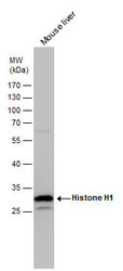
- Histone H1 antibody detects Histone H1 protein by western blot analysis. Mouse tissue extracts (50 ?g) was separated by 12% SDS-PAGE, and the membrane was blotted with Histone H1 antibody (GTX114462) diluted by 1:5000. The HRP-conjugated anti-rabbit IgG antibody (GTX213110-01) was used to detect the primary antibody.
- Submitted by
- GeneTex (provider)
- Main image
- Experimental details
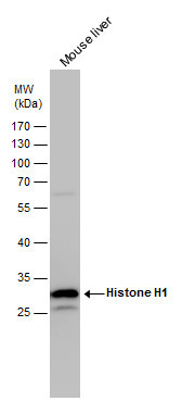
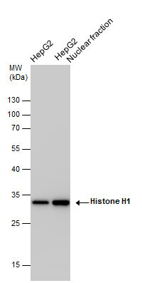
- Histone H1 antibody detects Histone H1 protein by western blot analysis. HepG2 whole cell extracts and nuclear extracts (30 ?g) were separated by 12% SDS-PAGE, and the membrane was blotted with Histone H1 antibody (GTX114462) at a dilution of 1:10000 and developed with Trident femto Western HRP Substrate (GTX14698). The HRP-conjugated anti-rabbit IgG antibody (GTX213110-01) was used to detect the primary antibody.
Supportive validation
- Submitted by
- GeneTex (provider)
- Main image
- Experimental details
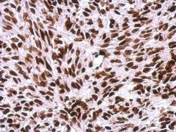
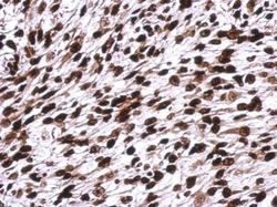
- Immunohistochemical analysis of paraffin-embedded SkHep1 xenograft, using Histone H1.0(GTX114462) antibody at 1:500 dilution.
- Submitted by
- GeneTex (provider)
- Main image
- Experimental details
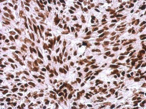
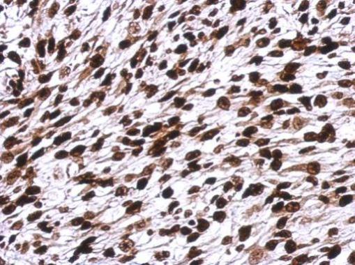
- Immunohistochemical analysis of paraffin-embedded C2C12 xenograft, using Histone H1.0(GTX114462) antibody at 1:500 dilution.
- Submitted by
- GeneTex (provider)
- Main image
- Experimental details
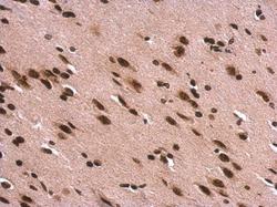
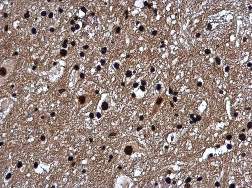
- Histone H1.0 antibody detects Histone H1.0 protein at nucleus on rat fore brain by immunohistochemical analysis. Sample: Paraffin-embedded rat fore brain. Histone H1.0 antibody (GTX114462) dilution: 1:500.
- Submitted by
- GeneTex (provider)
- Main image

- Experimental details
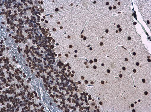
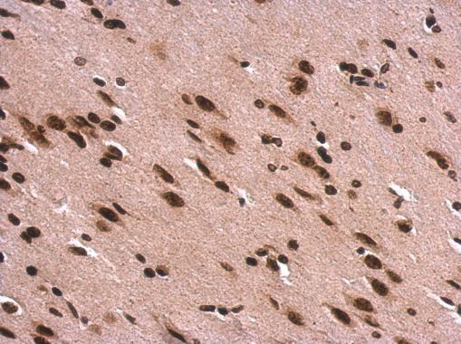
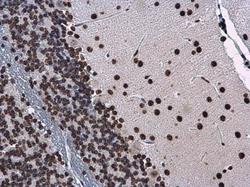
- Histone H1.0 antibody detects Histone H1.0 protein at nucleus in mouse brain by immunohistochemical analysis. Sample: Paraffin-embedded mouse brain. Histone H1.0 antibody (GTX114462) diluted at 1:500.
- Submitted by
- GeneTex (provider)
- Main image
- Experimental details
- Histone H1.0 antibody detects Histone H1.0 protein at nucleus in mouse brain by immunohistochemical analysis. Sample: Paraffin-embedded mouse brain. Histone H1.0 antibody (GTX114462) diluted at 1:500.