 Explore
 Validate
 Learn
Learn Western blot
 Immunocytochemistry
Antibody data
- Antibody Data
- Antigen structure
- References [0]
- Comments [0]
- Validations
- Immunocytochemistry [4]
- Immunohistochemistry [4]
- Chromatin Immunoprecipitation [2]
Submit
Validation data
Reference
Comment
Report error
- Product number
- PA5-51466 - Provider product page

- Provider
- Invitrogen Antibodies
- Product name
- Histone 1F0 Polyclonal Antibody
- Antibody type
- Polyclonal
- Antigen
- Recombinant protein fragment
- Description
- Immunogen sequence: MTENSTDHPK YSDMIVAAIQ AEKNRAGSSR QSIQKYIKSH YKVGENADSQ IKLSIKRLVT TGVLKQTKGV GASGSFRLAK SDEPKKSVAF KKTKKEIKKV ATPKKASK Highest antigen sequence identity to the following orthologs: Mouse - 93%, Rat - 39%.
- Reactivity
- Human
- Host
- Rabbit
- Isotype
- IgG
- Vial size
- 100 μL
- Concentration
- 0.05 mg/mL
- Storage
- Store at 4°C short term. For long term storage, store at -20°C, avoiding freeze/thaw cycles.
No comments: Submit comment
Supportive validation
- Submitted by
- Invitrogen Antibodies (provider)
- Main image
- Experimental details
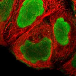
- Immunofluorescent staining of Histone 1F0 in human cell line A-431 using a Histone 1F0 Polyclonal Antibody (Product # PA5-51466) shows localization to nucleus and actin filaments.

- Submitted by
- Invitrogen Antibodies (provider)
- Main image

- Experimental details
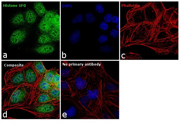
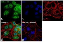
- Immunofluorescence analysis of Histone 1F0 was performed using 70% confluent log phase U2OS cells. The cells were fixed with 4% paraformaldehyde for 10 minutes, permeabilized with 0.1% Triton™ X-100 for 10 minutes, and blocked with 1% BSA for 1 hour at room temperature. The cells were labeled with Histone 1F0 Rabbit Polyclonal Antibody (Product # PA5-51466) at 5 microgram/mL in 0.1% BSA and incubated overnight at 4 degree and then labeled with Goat anti-Rabbit IgG (H+L) Superclonal™ Secondary Antibody, Alexa Fluor® 488 conjugate (Product # A27034) at a dilution of 1:2000 for 45 minutes at room temperature (Panel a: green). Nuclei (Panel b: blue) were stained with SlowFade® Gold Antifade Mountant with DAPI (Product # S36938). F-actin (Panel c: red) was stained with Rhodamine Phalloidin (Product # R415, 1:300). Panel d represents the merged image showing nuclear localization. Panel e represents control cells with no primary antibody to assess background. The images were captured at 60X magnification.
- Submitted by
- Invitrogen Antibodies (provider)
- Main image
- Experimental details
- Immunofluorescence analysis of Histone 1F0 was performed using 70% confluent log phase U2OS cells. The cells were fixed with 4% paraformaldehyde for 10 minutes, permeabilized with 0.1% Triton™ X-100 for 10 minutes, and blocked with 1% BSA for 1 hour at room temperature. The cells were labeled with Histone 1F0 Rabbit Polyclonal Antibody (Product # PA5-51466) at 5 microgram/mL in 0.1% BSA and incubated overnight at 4 degree and then labeled with Goat anti-Rabbit IgG (Heavy Chain) Superclonal™ Secondary Antibody, Alexa Fluor® 488 conjugate (Product # A27034) at a dilution of 1:2000 for 45 minutes at room temperature (Panel a: green). Nuclei (Panel b: blue) were stained with SlowFade® Gold Antifade Mountant with DAPI (Product # S36938). F-actin (Panel c: red) was stained with Rhodamine Phalloidin (Product # R415, 1:300). Panel d represents the merged image showing nuclear localization. Panel e represents control cells with no primary antibody to assess background. The images were captured at 60X magnification.

- Submitted by
- Invitrogen Antibodies (provider)
- Main image

- Experimental details
- Immunofluorescent staining of Histone 1F0 in human cell line A-431 using a Histone 1F0 Polyclonal Antibody (Product # PA5-51466) shows localization to nucleus and actin filaments.

Supportive validation
- Submitted by
- Invitrogen Antibodies (provider)
- Main image
- Experimental details
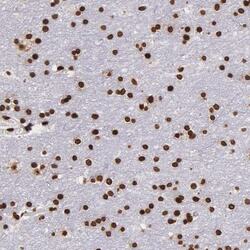
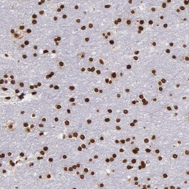
- Immunohistochemical analysis of Histone 1F0 in human cerebral cortex using Histone 1F0 Polyclonal Antibody (Product # PA5-51466) shows strong nuclear positivity in neurons.
- Submitted by
- Invitrogen Antibodies (provider)
- Main image

- Experimental details
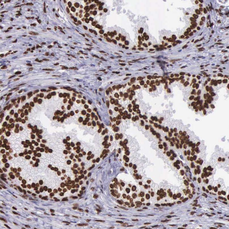
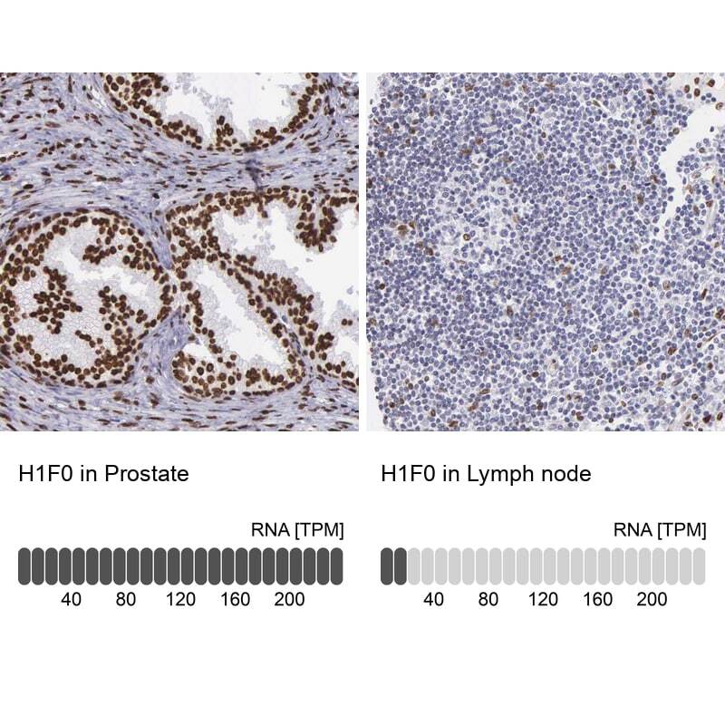
- Immunohistochemical staining of Histone 1F0 in human prostate using Histone 1F0 Polyclonal Antibody (Product # PA5-51466) shows high expression.
- Submitted by
- Invitrogen Antibodies (provider)
- Main image
- Experimental details
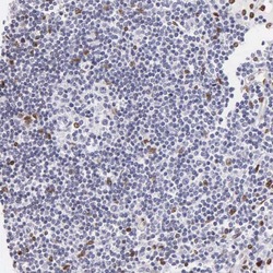
- Immunohistochemical staining of Histone 1F0 in human lymph node using Histone 1F0 Polyclonal Antibody (Product # PA5-51466) shows low expression as expected.
- Submitted by
- Invitrogen Antibodies (provider)
- Main image
- Experimental details
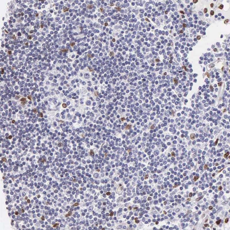
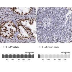
- Immunohistochemical staining of Histone 1F0 in human prostate and lymph node tissues using Histone 1F0 Polyclonal Antibody (Product # PA5-51466). Corresponding H1F0 RNA-seq data are presented for the same tissues.
Supportive validation
- Submitted by
- Invitrogen Antibodies (provider)
- Main image

- Experimental details
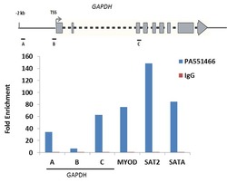
- Enrichment of endogenous Histone H2A.X protein at specific gene loci using Anti-Histone H2A.X Antibody: Chromatin Immunoprecipitation (ChIP) was performed using Anti-Histone H2A.X Polyclonal Antibody (Product # PA1-41004, 3 µg) on sheared chromatin from 2 million U-2 OS cells using the "MAGnify ChIP system" kit (Product # 49-2024). Normal Rabbit IgG was used as a negative IP control. The purified DNA was analyzed by qPCR with PCR primer pairs over the GAPDH gene (active) and MYOD, SAT2 satellite repeats, SAT Alpha (inactive). A schematic diagram of the GAPDH gene is shown on top of the figure. Data is presented as fold enrichment of the antibody signal versus the negative control IgG using the comparative CT method.

- Submitted by
- Invitrogen Antibodies (provider)
- Main image
- Experimental details
- Enrichment of endogenous Histone H2A.X protein at specific gene loci using Anti-Histone H2A.X Antibody: Chromatin Immunoprecipitation (ChIP) was performed using Anti-Histone H2A.X Polyclonal Antibody (Product # PA1-41004, 3 µg) on sheared chromatin from 2 million U-2 OS cells using the "MAGnify ChIP system" kit (Product # 49-2024). Normal Rabbit IgG was used as a negative IP control. The purified DNA was analyzed by qPCR with PCR primer pairs over the GAPDH gene (active) and MYOD, SAT2 satellite repeats, SAT Alpha (inactive). A schematic diagram of the GAPDH gene is shown on top of the figure. Data is presented as fold enrichment of the antibody signal versus the negative control IgG using the comparative CT method.
