Explore
 Validate
 Learn
Learn Western blot
Antibody data
- Antibody Data
- Antigen structure
- References [0]
- Comments [0]
- Validations
- Western blot [1]
- Immunohistochemistry [4]
Submit
Validation data
Reference
Comment
Report error
- Product number
- NBP1-83283 - Provider product page

- Provider
- Novus Biologicals
- Proper citation
- Novus Cat#NBP1-83283, RRID:AB_11021771
- Product name
- Rabbit Polyclonal TMEM176A Antibody
- Antibody type
- Polyclonal
- Description
- Immunogen affinity purified. Specificity of human TMEM176A antibody verified on a Protein Array containing target protein plus 383 other non-specific proteins.
- Reactivity
- Human
- Host
- Rabbit
- Isotype
- IgG
- Vial size
- 0.1 ml
- Storage
- Store at 4C short term. Aliquot and store at -20C long term. Avoid freeze-thaw cycles.
No comments: Submit comment
Supportive validation
- Submitted by
- Novus Biologicals (provider)
- Main image

- Experimental details
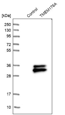
- Western Blot: TMEM176A Antibody [NBP1-83283] - Analysis in control (vector only transfected HEK293T lysate) and TMEM176A over-expression lysate (Co-expressed with a C-terminal myc-DDK tag (3.1 kDa) in mammalian HEK293T cells).
Supportive validation
- Submitted by
- Novus Biologicals (provider)
- Main image
- Experimental details
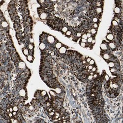
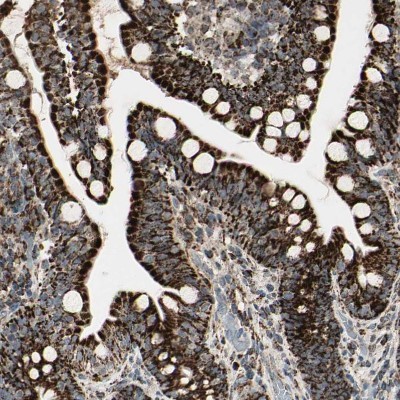
- Immunohistochemistry-Paraffin: TMEM176A Antibody [NBP1-83283] - Staining of human small intestine shows moderate to strong cytoplasmic positivity in glandular cells.
- Submitted by
- Novus Biologicals (provider)
- Main image

- Experimental details
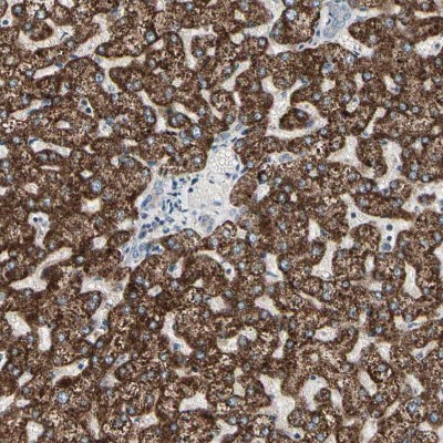
- Immunohistochemistry-Paraffin: TMEM176A Antibody [NBP1-83283] - Staining of human liver shows strong cytoplasmic positivity in hepatocytes.
- Submitted by
- Novus Biologicals (provider)
- Main image
- Experimental details
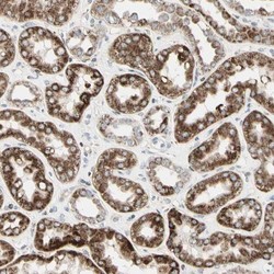
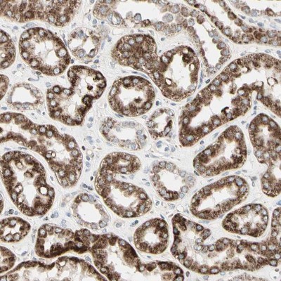
- Immunohistochemistry-Paraffin: TMEM176A Antibody [NBP1-83283] - Staining of human kidney shows strong cytoplasmic positivity in cells in tubules.
- Submitted by
- Novus Biologicals (provider)
- Main image

- Experimental details
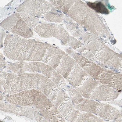
- Immunohistochemistry-Paraffin: TMEM176A Antibody [NBP1-83283] - Staining of human skeletal muscle shows no positivity in myocytes.