Explore
 Validate
 Learn
Learn Western blot
 Immunocytochemistry
Antibody data
- Antibody Data
- Antigen structure
- References [1]
- Comments [0]
- Validations
- Immunocytochemistry [1]
- Immunohistochemistry [1]
- Other assay [1]
Submit
Validation data
Reference
Comment
Report error
- Product number
- PA5-28534 - Provider product page

- Provider
- Invitrogen Antibodies
- Product name
- Latexin Polyclonal Antibody
- Antibody type
- Polyclonal
- Antigen
- Recombinant full-length protein
- Description
- Recommended positive controls: 293T, HeLaS3, Molt-4, mouse brain, rat brain, , rat heart. Predicted reactivity: Mouse (85%), Rat (85%), Rhesus Monkey (97%), Bovine (85%). Store product as a concentrated solution. Centrifuge briefly prior to opening the vial.
- Reactivity
- Human, Mouse, Rat
- Host
- Rabbit
- Isotype
- IgG
- Vial size
- 100 μL
- Concentration
- 1 mg/mL
- Storage
- Store at 4°C short term. For long term storage, store at -20°C, avoiding freeze/thaw cycles.
Submitted references Acute Sleep Loss Upregulates the Synaptic Scaffolding Protein, Homer1a, in Non-canonical Sleep/Wake Brain Regions, Claustrum, Piriform and Cingulate Cortices.
Zhu J, Hafycz J, Keenan BT, Guo X, Pack A, Naidoo N
Frontiers in neuroscience 2020;14:188
Frontiers in neuroscience 2020;14:188
No comments: Submit comment
Supportive validation
- Submitted by
- Invitrogen Antibodies (provider)
- Main image

- Experimental details
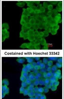
- Immunofluorescent analysis of LXN in paraformaldehyde-fixed mouse ESC D3 cells using a LXN polyclonal antibody (Product # PA5-28534) at a 1:200 dilution.
Supportive validation
- Submitted by
- Invitrogen Antibodies (provider)
- Main image
- Experimental details
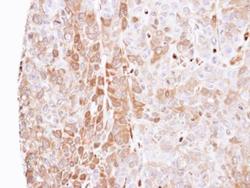
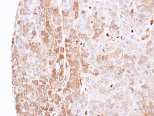
- Immunohistochemical analysis of paraffin-embedded FaDu xenograft, using LXN (Product # PA5-28534) antibody at 1:500 dilution. Antigen Retrieval: EDTA based buffer, pH 8.0, 15 min.
Supportive validation
- Submitted by
- Invitrogen Antibodies (provider)
- Main image
- Experimental details
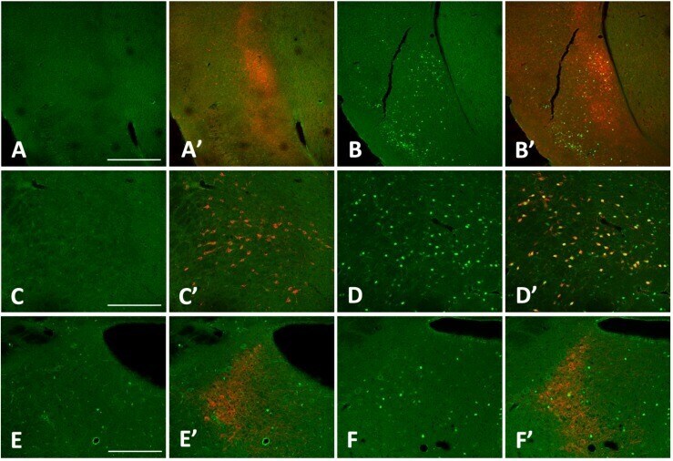
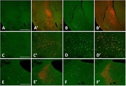
- FIGURE 5 cFos levels increase in the claustrum, lateral hypothalamus (LHA), and locus coeruleus (LC) with 3 h sleep deprivation (SD) compared to sleeping control (SC) animals. (A) 3 h SC cFos staining; (A') 3 h sleep control with cFos and Latexin as marker for the claustrum; (B) 3 h SD cFos staining; (B') 3 h SD with cFos and Latexin; (C) 3 h SC cFos staining; (C') 3 h sleep control with cFos and Orexin as marker for the LHA; (D) 3 h SD cFos staining; (D') 3 h SD with cFos and Orexin; (E) 3 h SC cFos staining; (E') 3 h sleep control with cFos and tyrosine hydroxylase (TH) as marker for the LC; F) 3 h SD cFos staining; (F') 3 h SD with cFos and TH. Bar size A: 500 mum; C,E : 250 mum.