Explore
 Validate
 Learn
Learn Western blot
 Immunocytochemistry
Antibody data
- Antibody Data
- Antigen structure
- References [2]
- Comments [0]
- Validations
- Immunocytochemistry [1]
Submit
Validation data
Reference
Comment
Report error
- Product number
- HPA011912 - Provider product page

- Provider
- Atlas Antibodies
- Proper citation
- Atlas Antibodies Cat#HPA011912, RRID:AB_1856277
- Product name
- Anti-PDLIM4
- Antibody type
- Polyclonal
- Description
- Polyclonal Antibody against Human PDLIM4, Gene description: PDZ and LIM domain 4, Alternative Gene Names: RIL, Validated applications: ICC, IHC, WB, Uniprot ID: P50479, Storage: Store at +4°C for short term storage. Long time storage is recommended at -20°C.
- Reactivity
- Human, Rat
- Host
- Rabbit
- Conjugate
- Unconjugated
- Isotype
- IgG
- Vial size
- 100 µl
- Concentration
- 0.1 mg/ml
- Storage
- Store at +4°C for short term storage. Long time storage is recommended at -20°C.
- Handling
- The antibody solution should be gently mixed before use.
Submitted references Prognostic Significance of EDN/RB, HJURP, p60/CAF-1 and PDLI4, Four New Markers in High-Grade Gliomas
Immunofluorescence and fluorescent-protein tagging show high correlation for protein localization in mammalian cells
Jiang T, de Tayrac M, Saikali S, Aubry M, Bellaud P, Boniface R, Quillien V, Mosser J
PLoS ONE 2013;8(9):e73332
PLoS ONE 2013;8(9):e73332
Immunofluorescence and fluorescent-protein tagging show high correlation for protein localization in mammalian cells
Stadler C, Rexhepaj E, Singan V, Murphy R, Pepperkok R, Uhlén M, Simpson J, Lundberg E
Nature Methods 2013;10(4):315-323
Nature Methods 2013;10(4):315-323
No comments: Submit comment
Supportive validation
- Submitted by
- Atlas Antibodies (provider)
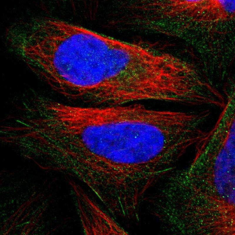
- Main image
- Experimental details
- Immunofluorescent staining of human cell line U-2 OS shows localization to actin filaments.
- Sample type
- Human