Explore
 Validate
 Learn
Learn Western blot
 ELISA
ELISAAntibody data
- Antibody Data
- Antigen structure
- References [5]
- Comments [0]
- Validations
- Western blot [1]
- Immunocytochemistry [1]
Submit
Validation data
Reference
Comment
Report error
- Product number
- 20541-1-AP - Provider product page

- Provider
- Proteintech Group
- Proper citation
- Proteintech Cat#20541-1-AP, RRID:AB_10694570
- Product name
- SH3BP1 antibody
- Antibody type
- Polyclonal
- Description
- SH3BP1 antibody (Cat. #20541-1-AP) is a rabbit polyclonal antibody that shows reactivity with human, mouse and has been validated for the following applications: IF, WB, ELISA.
- Reactivity
- Human, Mouse
- Host
- Rabbit
- Conjugate
- Unconjugated
- Isotype
- IgG
- Vial size
- 20ul, 150ul
Submitted references Exploring potential therapeutic targets for colorectal tumors based on whole genome sequencing of colorectal tumors and paracancerous tissues.
NicheProt: Cell-type-resolved proteomics of tissue compartments.
Prognostic impact of ARHGAP43(SH3BP1) in acute myeloid leukemia.
Reciprocal interactions among Cobll1, PACSIN2, and SH3BP1 regulate drug resistance in chronic myeloid leukemia.
Phosphoinositide 3-kinase enables phagocytosis of large particles by terminating actin assembly through Rac/Cdc42 GTPase-activating proteins.
Sheng Y, Niu S, Li D, Meng C, Wang T
Frontiers in molecular biosciences 2025;12:1605117
Frontiers in molecular biosciences 2025;12:1605117
NicheProt: Cell-type-resolved proteomics of tissue compartments.
Wu YC, Schwartz D, Abi Khalil E, Upadhye A, Rehman J, Lee SS
bioRxiv : the preprint server for biology 2025 Oct 24;
bioRxiv : the preprint server for biology 2025 Oct 24;
Prognostic impact of ARHGAP43(SH3BP1) in acute myeloid leukemia.
Yang L, Xu Q, Li J
Journal of the Formosan Medical Association = Taiwan yi zhi 2024 Sep;123(9):992-1003
Journal of the Formosan Medical Association = Taiwan yi zhi 2024 Sep;123(9):992-1003
Reciprocal interactions among Cobll1, PACSIN2, and SH3BP1 regulate drug resistance in chronic myeloid leukemia.
Park K, Yoo HS, Oh CK, Lee JR, Chung HJ, Kim HN, Kim SH, Kee KM, Kim TY, Kim M, Kim BG, Ra JS, Myung K, Kim H, Han SH, Seo MD, Lee Y, Kim DW
Cancer medicine 2022 Nov;11(21):4005-4020
Cancer medicine 2022 Nov;11(21):4005-4020
Phosphoinositide 3-kinase enables phagocytosis of large particles by terminating actin assembly through Rac/Cdc42 GTPase-activating proteins.
Schlam D, Bagshaw RD, Freeman SA, Collins RF, Pawson T, Fairn GD, Grinstein S
Nature communications 2015 Oct 14;6:8623
Nature communications 2015 Oct 14;6:8623
No comments: Submit comment
Supportive validation
- Submitted by
- Proteintech Group (provider)
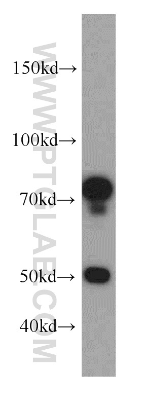
- Main image

- Experimental details
- mouse lung tissue were subjected to SDS PAGE followed by western blot with 20541-1-AP(SH3BP1 antibody) at dilution of 1:500
- Sample type
- tissue
Supportive validation
- Submitted by
- Proteintech Group (provider)
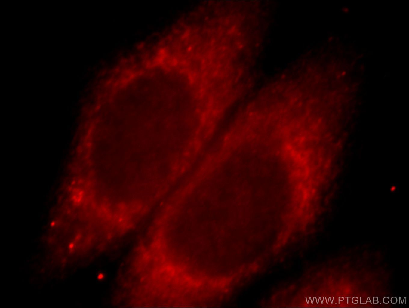
- Main image
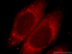
- Experimental details
- Immunofluorescent analysis of HepG2 cells, using SH3BP1 antibody 20541-AP at 1:25 dilution and Rhodamine-labeled goat anti-rabbit IgG (red).
- Sample type
- cell line