 Explore
 Validate
 Learn
Learn Western blot
 Immunocytochemistry
Antibody data
- Antibody Data
- Antigen structure
- References [1]
- Comments [0]
- Validations
- Immunocytochemistry [1]
Submit
Validation data
Reference
Comment
Report error
- Product number
- HPA009295 - Provider product page

- Provider
- Atlas Antibodies
- Proper citation
- Atlas Antibodies Cat#HPA009295, RRID:AB_1858425
- Product name
- Anti-TTC14
- Antibody type
- Polyclonal
- Description
- Polyclonal Antibody against Human TTC14, Gene description: tetratricopeptide repeat domain 14, Alternative Gene Names: FLJ00166, KIAA1980, Validated applications: WB, ICC, IHC, Uniprot ID: Q96N46, Storage: Store at +4°C for short term storage. Long time storage is recommended at -20°C.
- Reactivity
- Human, Mouse
- Host
- Rabbit
- Conjugate
- Unconjugated
- Isotype
- IgG
- Vial size
- 100 µl
- Concentration
- 0.05 mg/ml
- Storage
- Store at +4°C for short term storage. Long time storage is recommended at -20°C.
- Handling
- The antibody solution should be gently mixed before use.
Submitted references Unraveling transcriptome dynamics in human spermatogenesis.
Jan SZ, Vormer TL, Jongejan A, Röling MD, Silber SJ, de Rooij DG, Hamer G, Repping S, van Pelt AMM
Development (Cambridge, England) 2017 Oct 15;144(20):3659-3673
Development (Cambridge, England) 2017 Oct 15;144(20):3659-3673
No comments: Submit comment
Supportive validation
- Submitted by
- Atlas Antibodies (provider)
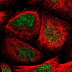
- Main image
- Experimental details
- Immunofluorescent staining of human cell line A-431 shows localization to nucleus.
- Sample type
- Human
