Explore
 Validate
 Learn
Learn Western blot
 Immunohistochemistry
Antibody data
- Antibody Data
- Antigen structure
- References [1]
- Comments [0]
- Validations
- Immunohistochemistry [2]
- Other assay [1]
Submit
Validation data
Reference
Comment
Report error
- Product number
- PA5-78512 - Provider product page

- Provider
- Invitrogen Antibodies
- Product name
- PARP14 Polyclonal Antibody
- Antibody type
- Polyclonal
- Antigen
- Recombinant full-length protein
- Description
- Positive Control: U87-MG, SK-N-SH, SK-N-AS Store product as a concentrated solution. Centrifuge briefly prior to opening the vial.
- Reactivity
- Human
- Host
- Rabbit
- Isotype
- IgG
- Vial size
- 100 μL
- Concentration
- 1.29 mg/mL
- Storage
- Store at 4°C short term. For long term storage, store at -20°C, avoiding freeze/thaw cycles.
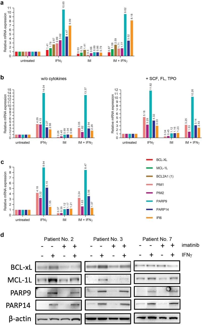
Submitted references IFNγ directly counteracts imatinib-induced apoptosis of primary human CD34+ CML stem/progenitor cells potentially through the upregulation of multiple key survival factors.
Ujvari D, Malyukova A, Zovko A, Yektaei-Karin E, Madapura HS, Keszei M, Nagy N, Lotfi K, Björn N, Wallvik J, Stenke L, Salamon D
Oncoimmunology 2022;11(1):2109861
Oncoimmunology 2022;11(1):2109861
No comments: Submit comment
Supportive validation
- Submitted by
- Invitrogen Antibodies (provider)
- Main image

- Experimental details
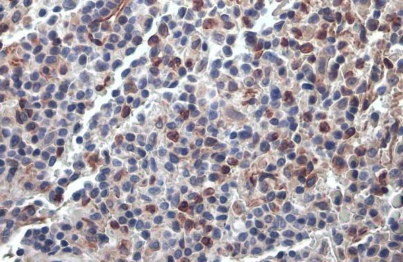
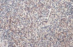
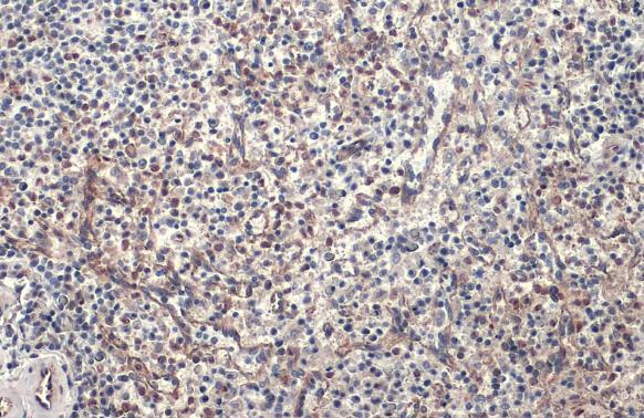
- PARP14 Polyclonal Antibody detects PARP14 protein at cell membrane and cytoplasm by immunohistochemical analysis. Sample: Paraffin-embedded human spleen. PARP14 stained by PARP14 Polyclonal Antibody (Product # PA5-78512) diluted at 1:500. Antigen Retrieval: Citrate buffer, pH 6.0, 15 min.
- Submitted by
- Invitrogen Antibodies (provider)
- Main image
- Experimental details
- PARP14 Polyclonal Antibody detects PARP14 protein at cell membrane and cytoplasm by immunohistochemical analysis. Sample: Paraffin-embedded human spleen. PARP14 stained by PARP14 Polyclonal Antibody (Product # PA5-78512) diluted at 1:500. Antigen Retrieval: Citrate buffer, pH 6.0, 15 min.
Supportive validation
- Submitted by
- Invitrogen Antibodies (provider)
- Main image
- Experimental details
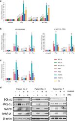
- Figure 7. IFNgamma upregulates the expression of several key anti-apoptotic genes in imatinib-treated primary human CD34+ CML stem/progenitor cells. (a) Relative mRNA levels of selected key anti-apoptotic genes normalized to EEF1A1, quantified by real-time RT-PCR in primary human CD34+ CML stem/progenitor cells (obtained from the peripheral blood of Patient No. 4) left untreated or treated for 18 hours with 5 muM imatinib (IM) and/or 5 ng/ml IFNgamma. (b) Relative mRNA levels of selected key anti-apoptotic genes normalized to EEF1A1, quantified by real-time RT-PCR in primary human CD34+ CML stem/progenitor cells (obtained from the peripheral blood of Patient No. 5) left untreated or treated for 90 minutes with 5 muM imatinib (IM) and/or 5 ng/ml IFNgamma in the absence (w/o cytokines) or presence of SCF, FL, and TPO. (c) Relative mRNA levels of selected key anti-apoptotic genes normalized to EEF1A1, quantified by real-time RT-PCR in primary human CD34+ CML stem/progenitor cells (obtained from the peripheral blood of Patient No. 6) left untreated or treated for 90 minutes with 5 muM imatinib (IM) and/or 5 ng/ml IFNgamma in the absence of cytokines. (d) Immunoblot analysis of BCL-XL, MCL-1L, PARP9, PARP14 and beta-actin protein expression in total cell extracts of CD34+ stem/progenitor cells (obtained from the peripheral blood of Patients Nos. 2, 3 and 7) left untreated (-) or treated (+) for 18 hours with 5 muM imatinib and/or 5 ng/ml IFNgamma. PIM1 and PIM2 proteins were expre