Antibody data
- Antibody Data
- Antigen structure
- References [9]
- Comments [0]
- Validations
- Immunocytochemistry [1]
Submit
Validation data
Reference
Comment
Report error
- Product number
- HPA012063 - Provider product page

- Provider
- Atlas Antibodies
- Proper citation
- Atlas Antibodies Cat#HPA012063, RRID:AB_1854981
- Product name
- Anti-PARP14
- Antibody type
- Polyclonal
- Description
- Polyclonal Antibody against Human PARP14, Gene description: poly (ADP-ribose) polymerase family, member 14, Alternative Gene Names: KIAA1268, pART8, Validated applications: IHC, ICC, Uniprot ID: Q460N5, Storage: Store at +4°C for short term storage. Long time storage is recommended at -20°C.
- Reactivity
- Human
- Host
- Rabbit
- Conjugate
- Unconjugated
- Isotype
- IgG
- Vial size
- 100 µl
- Concentration
- 0.1 mg/ml
- Storage
- Store at +4°C for short term storage. Long time storage is recommended at -20°C.
- Handling
- The antibody solution should be gently mixed before use.
Submitted references
PARP14 is regulated by the PARP9/DTX3L complex and promotes interferon γ-induced ADP-ribosylation
PARP14 is a novel target in STAT6 mutant follicular lymphoma
PARP14 inhibits microglial activation via LPAR5 to promote post-stroke functional recovery
The coronavirus macrodomain is required to prevent PARP-mediated inhibition of virus replication and enhancement of IFN expression
Identification of a novel biomarker in tangeretin‑induced cell death in AGS human gastric cancer cells
PARP14 promotes the Warburg effect in hepatocellular carcinoma by inhibiting JNK1-dependent PKM2 phosphorylation and activation
A systematic analysis of the PARP protein family identifies new functions critical for cell physiology
A systematic analysis of the PARP protein family identifies new functions critical for cell physiology
Parthasarathy S, Saenjamsai P, Hao H, Ferkul A, Pfannenstiel J, Bejan D, Chen Y, Suder E, Schwarting N, Aikawa M, Muhlberger E, Hume A, Orozco R, Sullivan C, Cohen M, Davido D, Fehr A
2025
2025
PARP14 is regulated by the PARP9/DTX3L complex and promotes interferon γ-induced ADP-ribosylation
Ribeiro V, Russo L, Hoch N
The EMBO Journal 2024;43(14):2908-2928
The EMBO Journal 2024;43(14):2908-2928
PARP14 is a novel target in STAT6 mutant follicular lymphoma
Mentz M, Keay W, Strobl C, Antoniolli M, Adolph L, Heide M, Lechner A, Haebe S, Osterode E, Kridel R, Ziegenhain C, Wange L, Hildebrand J, Shree T, Silkenstedt E, Staiger A, Ott G, Horn H, Szczepanowski M, Richter J, Levy R, Rosenwald A, Enard W, Zimber-Strobl U, von Bergwelt-Baildon M, Hiddemann W, Klapper W, Schmidt-Supprian M, Rudelius M, Bararia D, Passerini V, Weigert O
Leukemia 2022;36(9):2281-2292
Leukemia 2022;36(9):2281-2292
PARP14 inhibits microglial activation via LPAR5 to promote post-stroke functional recovery
Tang Y, Liu J, Wang Y, Yang L, Han B, Zhang Y, Bai Y, Shen L, Li M, Jiang T, Ye Q, Yu X, Huang R, Zhang Z, Xu Y, Yao H
Autophagy 2020;17(10):2905-2922
Autophagy 2020;17(10):2905-2922
The coronavirus macrodomain is required to prevent PARP-mediated inhibition of virus replication and enhancement of IFN expression
Weber F, Grunewald M, Chen Y, Kuny C, Maejima T, Lease R, Ferraris D, Aikawa M, Sullivan C, Perlman S, Fehr A
PLOS Pathogens 2019;15(5):e1007756
PLOS Pathogens 2019;15(5):e1007756
Identification of a novel biomarker in tangeretin‑induced cell death in AGS human gastric cancer cells
Yumnam S, Raha S, Kim S, Venkatarame Gowda Saralamma V, Lee H, Ha S, Heo J, Lee S, Kim E, Lee W, Kim J, Kim G
Oncology Reports 2018
Oncology Reports 2018
PARP14 promotes the Warburg effect in hepatocellular carcinoma by inhibiting JNK1-dependent PKM2 phosphorylation and activation
Iansante V, Choy P, Fung S, Liu Y, Chai J, Dyson J, Del Rio A, D’Santos C, Williams R, Chokshi S, Anders R, Bubici C, Papa S
Nature Communications 2015;6(1)
Nature Communications 2015;6(1)
A systematic analysis of the PARP protein family identifies new functions critical for cell physiology
Vyas S, Chesarone-Cataldo M, Todorova T, Huang Y, Chang P
Nature Communications 2013;4(1)
Nature Communications 2013;4(1)
A systematic analysis of the PARP protein family identifies new functions critical for cell physiology
Vyas S, Chesarone-Cataldo M, Todorova T, Huang Y, Chang P
Nature Communications 2013 August;4
Nature Communications 2013 August;4
No comments: Submit comment
Supportive validation
- Submitted by
- Atlas Antibodies (provider)
- Main image

- Experimental details
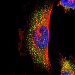
- Immunofluorescent staining of human cell line U-251 MG shows localization to cytosol.
- Sample type
- Human
 Explore
 Validate
 Learn
Learn Immunocytochemistry
 Immunohistochemistry