Explore
 Validate
 Learn
LearnAF744
antibody from Novus Biologicals
Targeting: TNFSF15
MGC129934, MGC129935, TL1, TL1A, VEGI, VEGI192A
 Western blot
 Immunohistochemistry
Antibody data
- Antibody Data
- Antigen structure
- References [1]
- Comments [0]
- Validations
- Immunohistochemistry [1]
Submit
Validation data
Reference
Comment
Report error
- Product number
- AF744 - Provider product page

- Provider
- Novus Biologicals
- Product name
- Goat Polyclonal TL1A/TNFSF15 Antibody
- Antibody type
- Polyclonal
- Description
- Antigen Affinity-purified. Detects human TL1A in direct ELISAs and Western blots. In direct ELISAs and Western blots, approximately 5% cross-reactivity with recombinant mouse TL1A is observed, and less than 2% cross-reactivity with recombinant human (rh) APRIL, rhTNF-alpha , and rhFas Ligand is observed.
- Reactivity
- Human
- Host
- Goat
- Conjugate
- Unconjugated
- Isotype
- IgG
- Vial size
- 100 ug
- Concentration
- LYOPH
- Storage
- Use a manual defrost freezer and avoid repeated freeze-thaw cycles. 12 months from date of receipt, -20 to -70 degreesC as supplied. 1 month, 2 to 8 degreesC under sterile conditions after reconstitution. 6 months, -20 to -70 degreesC under sterile conditions after reconstitution.
Submitted references The somatostatin analogue octreotide inhibits growth of small intestine neuroendocrine tumour cells.
Li SC, Martijn C, Cui T, Essaghir A, Luque RM, Demoulin JB, Castaño JP, Öberg K, Giandomenico V
PloS one 2012;7(10):e48411
PloS one 2012;7(10):e48411
No comments: Submit comment
Supportive validation
- Submitted by
- Novus Biologicals (provider)
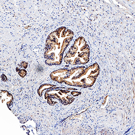
- Main image

- Experimental details
- TL1A/TNFSF15 in Human Prostate. TL1A/TNFSF15 was detected in immersion fixed paraffin-embedded sections of human prostate using Goat Anti-Human TL1A/TNFSF15 Antigen Affinity-purified Polyclonal Antibody (Catalog # AF744) at 3 µg/mL for 1 hour at room temperature followed by incubation with the Anti-Goat IgG VisUCyte™ HRP Polymer Antibody (Catalog # VC004). Tissue was stained using DAB (brown) and counterstained with hematoxylin (blue). Specific staining was localized to epithelial cells. View our protocol for IHC Staining with VisUCyte HRP Polymer Detection Reagents.