Explore
 Validate
 Learn
LearnMAB74421-100
antibody from Novus Biologicals
Targeting: TNFSF15
MGC129934, MGC129935, TL1, TL1A, VEGI, VEGI192A
 Western blot
Antibody data
- Antibody Data
- Antigen structure
- References [0]
- Comments [0]
- Validations
- Western blot [1]
- Immunohistochemistry [1]
- Blocking/Neutralizing [1]
Submit
Validation data
Reference
Comment
Report error
- Product number
- MAB74421-100 - Provider product page

- Provider
- Novus Biologicals
- Product name
- Rabbit Monoclonal TL1A/TNFSF15 Antibody
- Antibody type
- Monoclonal
- Description
- Protein A or G purified from cell culture supernatant. Detects human TL1A/TNFSF15 in direct ELISAs and Western blots.
- Reactivity
- Human
- Host
- Rabbit
- Conjugate
- Unconjugated
- Isotype
- IgG
- Vial size
- 100 ug
- Storage
- Use a manual defrost freezer and avoid repeated freeze-thaw cycles. 12 months from date of receipt, -20 to -70 degreesC as supplied. 1 month, 2 to 8 degreesC under sterile conditions after reconstitution. 6 months, -20 to -70 degreesC under sterile conditions after reconstitution.
No comments: Submit comment
Supportive validation
- Submitted by
- Novus Biologicals (provider)
- Main image
- Experimental details
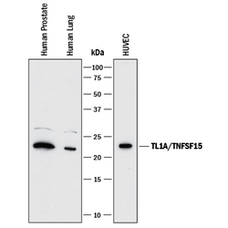
- Detection of Human TL1A/TNFSF15 by Western Blot. Western blot shows lysates of human prostate tissue, human lung tissue, and HUVEC human umbilical vein endothelial cells. PVDF membrane was probed with 1 µg/mL of Rabbit Anti-Human TL1A/ TNFSF15 Monoclonal Antibody (Catalog # MAB74421) followed by HRP-conjugated Anti-Rabbit IgG Secondary Antibody (Catalog # HAF008). A specific band was detected for TL1A/TNFSF15 at approximately 22 kDa (as indicated). This experiment was conducted under reducing conditions and using Immunoblot Buffer Group 1.
Supportive validation
- Submitted by
- Novus Biologicals (provider)
- Main image
- Experimental details
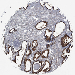
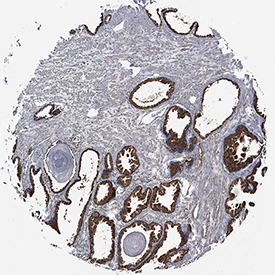
- TL1A/TNFSF15 in Human Prostate Cancer Tissue. TL1A/TNFSF15 was detected in immersion fixed paraffin-embedded sections of human prostate cancer tissue using Rabbit Anti-Human TL1A/TNFSF15 Monoclonal Antibody (Catalog # MAB74421) at 0.3 µg/mL for 1 hour at room temperature followed by incubation with the Anti-Mouse IgG VisUCyte™ HRP Polymer Antibody (Catalog # VC001). Tissue was stained using DAB (brown) and counterstained with hematoxylin (blue). Specific staining was localized to cytoplasm of epithelial cells. View our protocol for IHC Staining with VisUCyte HRP Polymer Detection Reagents.
Supportive validation
- Submitted by
- Novus Biologicals (provider)
- Main image
- Experimental details
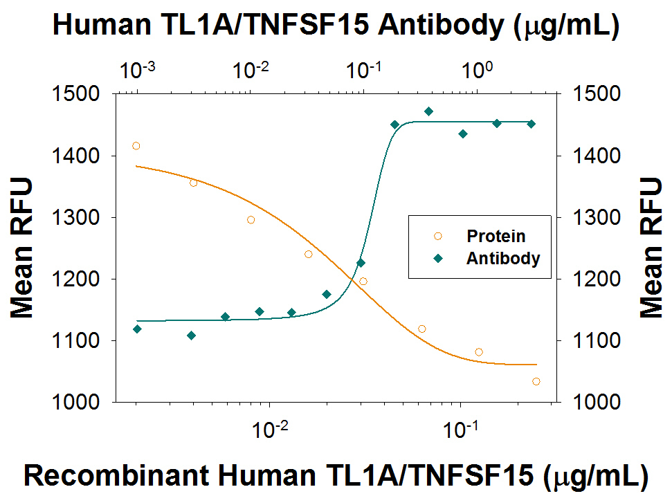
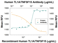
- Apoptosis Induced by TL1A/TNFSF15 and Neutralization by Human TL1A/TNFSF15 Antibody. Recombinant Human TL1A/TNFSF15 (Catalog # 1319-TL) induces apoptosis in the TF-1 human erythroleukemic cell line in a dose-dependent manner (orange line), as measured by Resazurin (Catalog # AR002). Apoptosis elicited by Recombinant Human TL1A/TNFSF15 (80 ng/mL) is neutralized (green line) by increasing concentrations of Rabbit Anti-Human TL1A/ TNFSF15 Monoclonal Antibody (Catalog # MAB74421). The ND50 is typically 0.04-0.2 ug/mL.