 Explore
 Validate
 Learn
Learn46-7911-82
antibody from Invitrogen Antibodies
Targeting: TNFSF15
MGC129934, MGC129935, TL1, TL1A, VEGI, VEGI192A
 Flow cytometry
Antibody data
- Antibody Data
- Antigen structure
- References [5]
- Comments [0]
- Validations
- Flow cytometry [1]
Submit
Validation data
Reference
Comment
Report error
- Product number
- 46-7911-82 - Provider product page

- Provider
- Invitrogen Antibodies
- Product name
- TL1A Monoclonal Antibody (Tandys1a), PerCP-eFluor™ 710, eBioscience™
- Antibody type
- Monoclonal
- Antigen
- Recombinant full-length protein
- Description
- Description: The Tandys1a monoclonal antibody reacts with human and mouse TL1A (TNFSF15). TL1A is a member of the TNF ligand family and is expressed by endothelial cells, tissue macrophages, monocyte-derived dendritic cells, plasma cells and lamina propria-derived lymphocytes. Among peripheral blood cells, TL1A has been shown to be expressed predominantly by CD4+CCR9+ lymphocytes. Its expression has been shown to be induced by TNF alpha, IL-1 alpha and IFN gamma. TL1A binds DR3, which has been shown to be specifically upregulated in Th17 cells among CD4+ T cells. TL1A-deficient dendritic cells are weak in their ability to support Th17 cells in vitro and show decreased severity of EAE. In vivo, TL1A has been shown to be upregulated in tissue lymphocytes of Crohn's Disease and ulcerative colitis patients. Applications Reported: This Tandys1a antibody has been reported for use in flow cytometric analysis, and intracellular staining followed by flow cytometric analysis. Applications Tested: This Tandys1a antibody has been tested by flow cytometric analysis of normal human peripheral blood cells or mouse splenocytes. This can be used at less than or equal to 0.5 µg per test. A test is defined as the amount (µg) of antibody that will stain a cell sample in a final volume of 100 µL. Cell number should be determined empirically but can range from 10^5 to 10^8 cells/test. It is recommended that the antibody be carefully titrated for optimal performance in the assay of interest. PerCP-eFluor® 710 can be used in place of PE-Cy5, PE-Cy5.5 or PerCP-Cy5.5. PerCP-eFluor® 710 emits at 710 nm and is excited with the blue laser (488 nm). Please make sure that your instrument is capable of detecting this fluorochrome. For a filter configuration, we recommend using the 685 LP dichroic mirror and 710/40 band pass filter, however the 695/40 band pass filter is an acceptable alternative. Our testing indicates that PerCP-eFluor® 710 conjugated antibodies are stable when stained samples are exposed to freshly prepared 2% formaldehyde overnight at 4°C, but please evaluate for alternative fixation protocols. Excitation: 488 nm; Emission: 710 nm; Laser: Blue Laser. Filtration: 0.2 µm post-manufacturing filtered.
- Reactivity
- Human, Mouse
- Host
- Mouse
- Isotype
- IgG
- Antibody clone number
- Tandys1a
- Vial size
- 100 µg
- Concentration
- 0.2 mg/mL
- Storage
- 4° C, store in dark, DO NOT FREEZE!
Submitted references TNF-like ligand 1A (TL1A) gene knockout leads to ameliorated collagen-induced arthritis in mice: implication of TL1A in humoral immune responses.
Naive and activated T cells display differential responsiveness to TL1A that affects Th17 generation, maintenance, and proliferation.
TL1A produced by lamina propria macrophages induces Th1 and Th17 immune responses in cooperation with IL-23 in patients with Crohn's disease.
The T cell costimulator TL1A is induced by FcgammaR signaling in human monocytes and dendritic cells.
Dominant role for TL1A/DR3 pathway in IL-12 plus IL-18-induced IFN-gamma production by peripheral blood and mucosal CCR9+ T lymphocytes.
Wang X, Hu Y, Charpentier T, Lamarre A, Qi S, Wu J, Luo H
Journal of immunology (Baltimore, Md. : 1950) 2013 Dec 1;191(11):5420-9
Journal of immunology (Baltimore, Md. : 1950) 2013 Dec 1;191(11):5420-9
Naive and activated T cells display differential responsiveness to TL1A that affects Th17 generation, maintenance, and proliferation.
Jones GW, Stumhofer JS, Foster T, Twohig JP, Hertzog P, Topley N, Williams AS, Hunter CA, Jenkins BJ, Wang EC, Jones SA
FASEB journal : official publication of the Federation of American Societies for Experimental Biology 2011 Jan;25(1):409-19
FASEB journal : official publication of the Federation of American Societies for Experimental Biology 2011 Jan;25(1):409-19
TL1A produced by lamina propria macrophages induces Th1 and Th17 immune responses in cooperation with IL-23 in patients with Crohn's disease.
Kamada N, Hisamatsu T, Honda H, Kobayashi T, Chinen H, Takayama T, Kitazume MT, Okamoto S, Koganei K, Sugita A, Kanai T, Hibi T
Inflammatory bowel diseases 2010 Apr;16(4):568-75
Inflammatory bowel diseases 2010 Apr;16(4):568-75
The T cell costimulator TL1A is induced by FcgammaR signaling in human monocytes and dendritic cells.
Prehn JL, Thomas LS, Landers CJ, Yu QT, Michelsen KS, Targan SR
Journal of immunology (Baltimore, Md. : 1950) 2007 Apr 1;178(7):4033-8
Journal of immunology (Baltimore, Md. : 1950) 2007 Apr 1;178(7):4033-8
Dominant role for TL1A/DR3 pathway in IL-12 plus IL-18-induced IFN-gamma production by peripheral blood and mucosal CCR9+ T lymphocytes.
Papadakis KA, Zhu D, Prehn JL, Landers C, Avanesyan A, Lafkas G, Targan SR
Journal of immunology (Baltimore, Md. : 1950) 2005 Apr 15;174(8):4985-90
Journal of immunology (Baltimore, Md. : 1950) 2005 Apr 15;174(8):4985-90
No comments: Submit comment
Supportive validation
- Submitted by
- Invitrogen Antibodies (provider)
- Main image
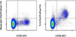
- Experimental details
- Normal human peripheral blood cells were stained with Fixable Viability Dye (FVD) eFluor® 780 (Product # 65-0865-14), then washed and stained with Anti-Human CD4, Anti-Human CCR9, and 0.25 µg of Mouse IgG1 K Isotype Control PerCP-eFluor® 710 (Product # 46-4714-82) (left) or 0.25 µg of Anti-Human/Mouse TL1A PerCP-eFluor® 710 (right). Viable CD4+ lymphocytes were used for analysis.
