Explore
 Validate
 Learn
LearnMAB74422-100
antibody from R&D Systems
Targeting: TNFSF15
MGC129934, MGC129935, TL1, TL1A, VEGI, VEGI192A
 Western blot
Antibody data
- Antibody Data
- Antigen structure
- References [0]
- Comments [0]
- Validations
- Western blot [1]
- Immunohistochemistry [1]
- Flow cytometry [1]
- Blocking/Neutralizing [1]
Submit
Validation data
Reference
Comment
Report error
- Product number
- MAB74422-100 - Provider product page

- Provider
- R&D Systems
- Product name
- Human TL1A/TNFSF15 Antibody
- Antibody type
- Monoclonal
- Description
- Protein A or G purified from cell culture supernatant. Detects human TL1A/TNFSF15 in direct ELISAs.
- Reactivity
- Human
- Host
- Rabbit
- Conjugate
- Unconjugated
- Antigen sequence
O95150- Isotype
- IgG
- Antibody clone number
- 2116A
- Vial size
- 100 ug
- Storage
- Use a manual defrost freezer and avoid repeated freeze-thaw cycles. 12 months from date of receipt, -20 to -70 °C as supplied. 1 month, 2 to 8 °C under sterile conditions after reconstitution. 6 months, -20 to -70 °C under sterile conditions after reconstitution.
No comments: Submit comment
Supportive validation
- Submitted by
- R&D Systems (provider)
- Main image
- Experimental details
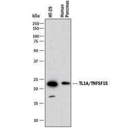
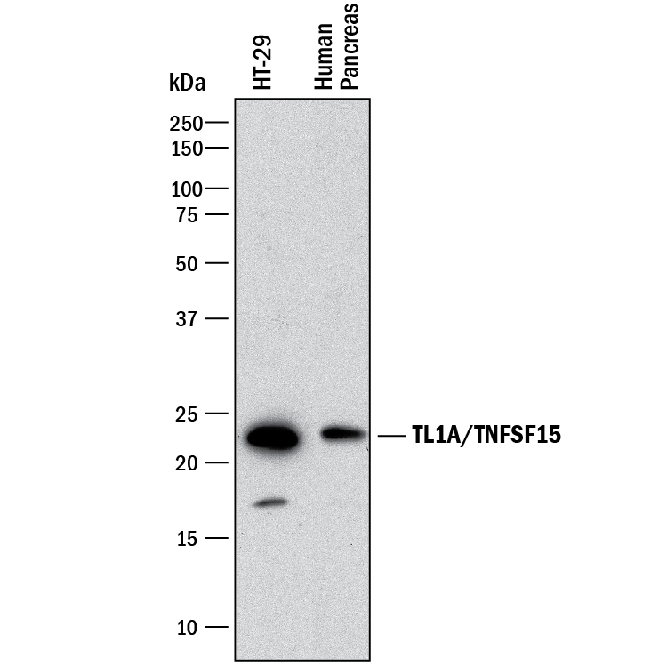
- Detection of Human TL1A/TNFSF15 by Western Blot. Western blot shows lysates of HT-29 human colon adenocarcinoma cell line and human pancreas tissue. PVDF membrane was probed with 1 µg/mL of Rabbit Anti-Human TL1A/TNFSF15 Monoclonal Antibody (Catalog # MAB74422) followed by HRP-conjugated Anti-Rabbit IgG Secondary Antibody (Catalog # HAF008). A specific band was detected for TL1A/TNFSF15 at approximately 22 kDa (as indicated). This experiment was conducted under reducing conditions and using Immunoblot Buffer Group 1.
Supportive validation
- Submitted by
- R&D Systems (provider)
- Main image
- Experimental details
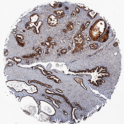
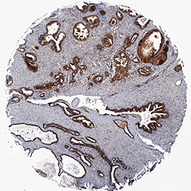
- TL1A/TNFSF15 in Human Prostate Cancer Tissue. TL1A/TNFSF15 was detected in immersion fixed paraffin-embedded sections of human prostate cancer tissue using Rabbit Anti-Human TL1A/TNFSF15 Monoclonal Antibody (Catalog # MAB74422) at 0.3 µg/mL for 1 hour at room temperature followed by incubation with the Anti-Rabbit IgG VisUCyte™ HRP Polymer Antibody (Catalog # VC003). Before incubation with the primary antibody, tissue was subjected to heat-induced epitope retrieval using Antigen Retrieval Reagent-Basic (Catalog # CTS013). Tissue was stained using DAB (brown) and counterstained with hematoxylin (blue). Specific staining was localized to cytoplasm in epithelial cells. View our protocol for IHC Staining with VisUCyte HRP Polymer Detection Reagents.
Supportive validation
- Submitted by
- R&D Systems (provider)
- Main image

- Experimental details
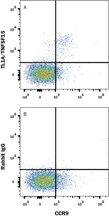
- Detection of TL1A/TNFSF15 in Human PBMCs by Flow Cytometry. Human peripheral blood mononuclear cells (PBMCs) were stained with (A) Rabbit Anti-Human TL1A/TNFSF15 Monoclonal Antibody (Catalog # MAB74422) or (B) Rabbit IgG control antibody (Catalog # MAB1050) followed by Goat anti-Rabbit IgG PE-conjugated Secondary Antibody (Catalog # F0110) and Mouse anti-Human CCR9 APC-conjugated Monoclonal Antibody (Catalog # FAB1791A). Cells were gated on CD4+ lymphocytes using Mouse anti-Human CD4 FITC-conjugated Monoclonal Antibody (Catalog # FAB3791F). View our protocol for Staining Membrane-associated Proteins.
Supportive validation
- Submitted by
- R&D Systems (provider)
- Main image
- Experimental details
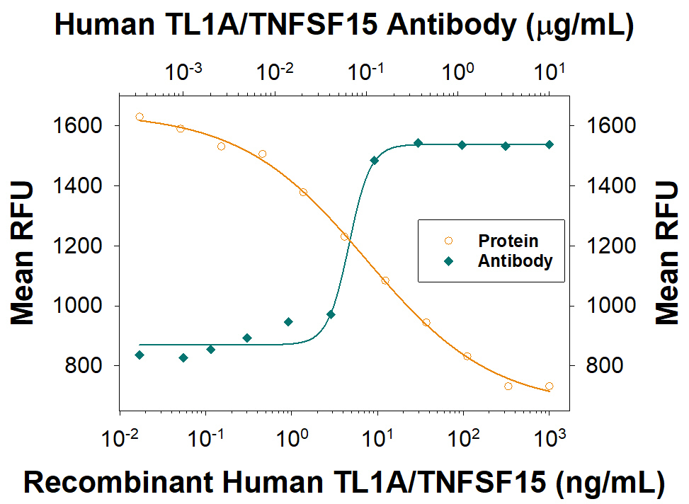
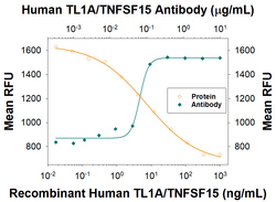
- Apoptosis Induced by TL1A/TNFSF15 and Neutralization by Human TL1A/TNFSF15 Antibody. Recombinant Human TL1A/TNFSF15 (Catalog # 1319-TL ) induces apoptosis in the TF-1 human erythroleukemic cell line in a dose-dependent manner (orange line), as measured by Resazurin (Catalog # AR002). Apoptosis elicited by Recombinant Human TL1A/TNFSF15 (80 ng/mL) is neutralized (green line) by increasing concentrations of Rabbit Anti-Human TL1A/TNFSF15 Monoclonal Antibody (Catalog # MAB74422). The ND50 is typically 0.04-0.2 μg/mL.