 Explore
 Validate
 Learn
Learn Immunocytochemistry
Antibody data
- Antibody Data
- Antigen structure
- References [0]
- Comments [0]
- Validations
- Immunocytochemistry [1]
- Immunohistochemistry [1]
- Flow cytometry [1]
Submit
Validation data
Reference
Comment
Report error
- Product number
- MAB52412-100 - Provider product page

- Provider
- R&D Systems
- Product name
- Mouse GITR/TNFRSF18 Antibody
- Antibody type
- Monoclonal
- Description
- Protein A or G purified from cell culture supernatant. Detects mouse GITR/TNFRSF18 in direct ELISAs.
- Reactivity
- Mouse
- Host
- Rabbit
- Conjugate
- Unconjugated
- Antigen sequence
O35714- Isotype
- IgG
- Antibody clone number
- 2375B
- Vial size
- 100 ug
- Storage
- Use a manual defrost freezer and avoid repeated freeze-thaw cycles. 12 months from date of receipt, -20 to -70 °C as supplied. 1 month, 2 to 8 °C under sterile conditions after reconstitution. 6 months, -20 to -70 °C under sterile conditions after reconstitution.
No comments: Submit comment
Supportive validation
- Submitted by
- R&D Systems (provider)
- Main image
- Experimental details
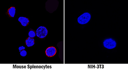
- GITR/TNFRSF18 in Mouse Splenocytes and NIH-3T3 Cell Line. GITR/TNFRSF18 was detected in immersion fixed mouse splenocytes (left panel; positive staining) and NIH-3T3 mouse embryonic fibroblast cell line (right panel; negative staining) using Rabbit Anti-Mouse GITR/TNFRSF18 Monoclonal Antibody (Catalog # MAB52412) at 8 µg/mL for 3 hours at room temperature. Cells were stained using the NorthernLights™ 557-conjugated Anti-Rabbit IgG Secondary Antibody (red; Catalog # NL004) and counterstained with DAPI (blue). Specific staining was localized to cell surfaces. View our protocol for Fluorescent ICC Staining of Non-adherent Cells.

Supportive validation
- Submitted by
- R&D Systems (provider)
- Main image

- Experimental details
- GITR/TNFRSF18 in Mouse Spleen. GITR/TNFRSF18 was detected in immersion fixed frozen sections of mouse spleen using Rabbit Anti-Mouse GITR/TNFRSF18 Monoclonal Antibody (Catalog # MAB52412) at 3 µg/mL for 1 hour at room temperature followed by incubation with the Anti-Rabbit IgG VisUCyte™ HRP Polymer Antibody (Catalog # VC003). Tissue was stained using DAB (brown) and counterstained with hematoxylin (blue). Specific staining was localized to lymphocytes. View our protocol for IHC Staining with VisUCyte HRP Polymer Detection Reagents.

Supportive validation
- Submitted by
- R&D Systems (provider)
- Main image
- Experimental details
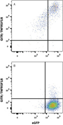
- Detection of GITR/TNFRSF18 in HEK293 Human Cell Line Transfected with Mouse GITR/TNFRSF18 and eGFP by Flow Cytometry. HEK293 human embryonic kidney cell line transfected with (A) mouse GITR/TNFRSF18 or (B) irrelevant transfectants and eGFP was stained with Rabbit Anti-Mouse GITR/TNFRSF18 Monoclonal Antibody (Catalog # MAB52412) followed by APC-conjugated Anti-Rabbit IgG Secondary Antibody (Catalog # F0111). Quadrant markers were set based on control antibody staining (Catalog # MAB1050). View our protocol for Staining Membrane-associated Proteins.
