Explore
 Validate
 Learn
Learn Western blot
Antibody data
- Antibody Data
- Antigen structure
- References [2]
- Comments [0]
- Validations
- Western blot [1]
- Immunohistochemistry [3]
- Flow cytometry [2]
- Other assay [4]
Submit
Validation data
Reference
Comment
Report error
- Product number
- PA5-26288 - Provider product page

- Provider
- Invitrogen Antibodies
- Product name
- SNRPD3 Polyclonal Antibody
- Antibody type
- Polyclonal
- Antigen
- Synthetic peptide
- Description
- This antibody is predicted to react with mouse and Xenopus based on sequence homology.
- Reactivity
- Human
- Host
- Rabbit
- Isotype
- IgG
- Vial size
- 400 μL
- Concentration
- 0.5 mg/mL
- Storage
- Store at 4°C short term. For long term storage, store at -20°C, avoiding freeze/thaw cycles.
Submitted references A missense mutation in SNRPE linked to non-syndromal microcephaly interferes with U snRNP assembly and pre-mRNA splicing.
Impaired spliceosomal UsnRNP assembly leads to Sm mRNA down-regulation and Sm protein degradation.
Chen T, Zhang B, Ziegenhals T, Prusty AB, Fröhler S, Grimm C, Hu Y, Schaefke B, Fang L, Zhang M, Kraemer N, Kaindl AM, Fischer U, Chen W
PLoS genetics 2019 Oct;15(10):e1008460
PLoS genetics 2019 Oct;15(10):e1008460
Impaired spliceosomal UsnRNP assembly leads to Sm mRNA down-regulation and Sm protein degradation.
Prusty AB, Meduri R, Prusty BK, Vanselow J, Schlosser A, Fischer U
The Journal of cell biology 2017 Aug 7;216(8):2391-2407
The Journal of cell biology 2017 Aug 7;216(8):2391-2407
No comments: Submit comment
Supportive validation
- Submitted by
- Invitrogen Antibodies (provider)
- Main image

- Experimental details
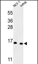
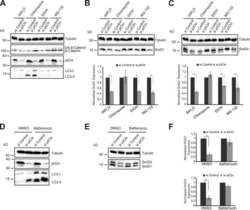
- Western blot analysis of SNRPD3 in MCF-7, Jurkat cell line lysates. Samples were incubated with SNRPD3 polyclonal antibody (Product # PA5-26288). Lysates: 35 µg/lane. SNRPD3 (arrow).
Supportive validation
- Submitted by
- Invitrogen Antibodies (provider)
- Main image
- Experimental details
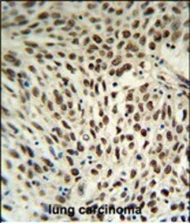
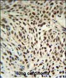
- Immunohistochemistry analysis of SNRPD3 in formalin fixed and paraffin embedded human lung carcinoma. Samples were incubated with SNRPD3 polyclonal antibody (Product # PA5-26288) followed by peroxidase conjugation of the secondary antibody and DAB staining. This data demonstrates the use of this antibody for immunohistochemistry. Clinical relevance has not been evaluated.
- Submitted by
- Invitrogen Antibodies (provider)
- Main image
- Experimental details
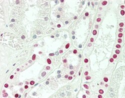
- Immunohistochemistry analysis of SNRPD3 in formalin-fixed and paraffin-embedded human kidney tissue. Samples were incubated with SNRPD3 polyclonal antibody (Product # PA5-26288). This data demonstrates the use of this antibody for immunohistochemistry. Clinical relevance has not been evaluated.

- Submitted by
- Invitrogen Antibodies (provider)
- Main image
- Experimental details
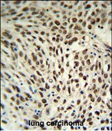
- Immunohistochemistry analysis of SNRPD3 in formalin fixed and paraffin embedded human lung carcinoma. Samples were incubated with SNRPD3 polyclonal antibody (Product # PA5-26288) followed by peroxidase conjugation of the secondary antibody and DAB staining. This data demonstrates the use of this antibody for immunohistochemistry. Clinical relevance has not been evaluated.

Supportive validation
- Submitted by
- Invitrogen Antibodies (provider)
- Main image
- Experimental details
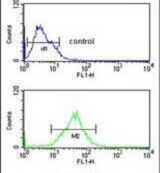
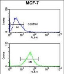
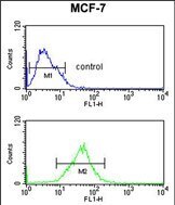
- Flow cytometry analysis of MCF-7 cells using a SNRPD3 polyclonal antibody (Product # PA5-26288) (bottom) compared to a negative control cell (top) at a dilution of 1:10-50, followed by a FITC-conjugated goat anti-rabbit antibody
- Submitted by
- Invitrogen Antibodies (provider)
- Main image
- Experimental details
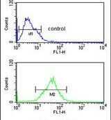
- Flow cytometry of SNRPD3 in MCF-7 cells (bottom histogram). Samples were incubated with SNRPD3 polyclonal antibody (Product # PA5-26288) followed by FITC-conjugated goat-anti-rabbit secondary antibody. Negative control cell (top histogram).
Supportive validation
- Submitted by
- Invitrogen Antibodies (provider)
- Main image
- Experimental details
- NULL
- Submitted by
- Invitrogen Antibodies (provider)
- Main image
- Experimental details
- NULL
- Submitted by
- Invitrogen Antibodies (provider)
- Main image
- Experimental details
- NULL

- Submitted by
- Invitrogen Antibodies (provider)
- Main image
- Experimental details
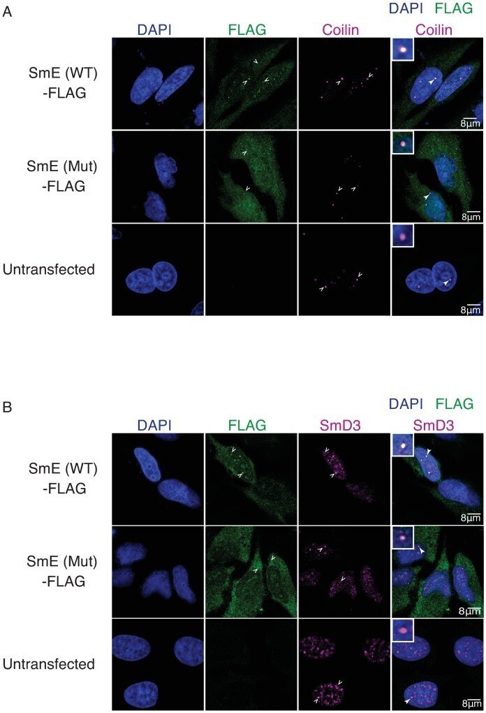
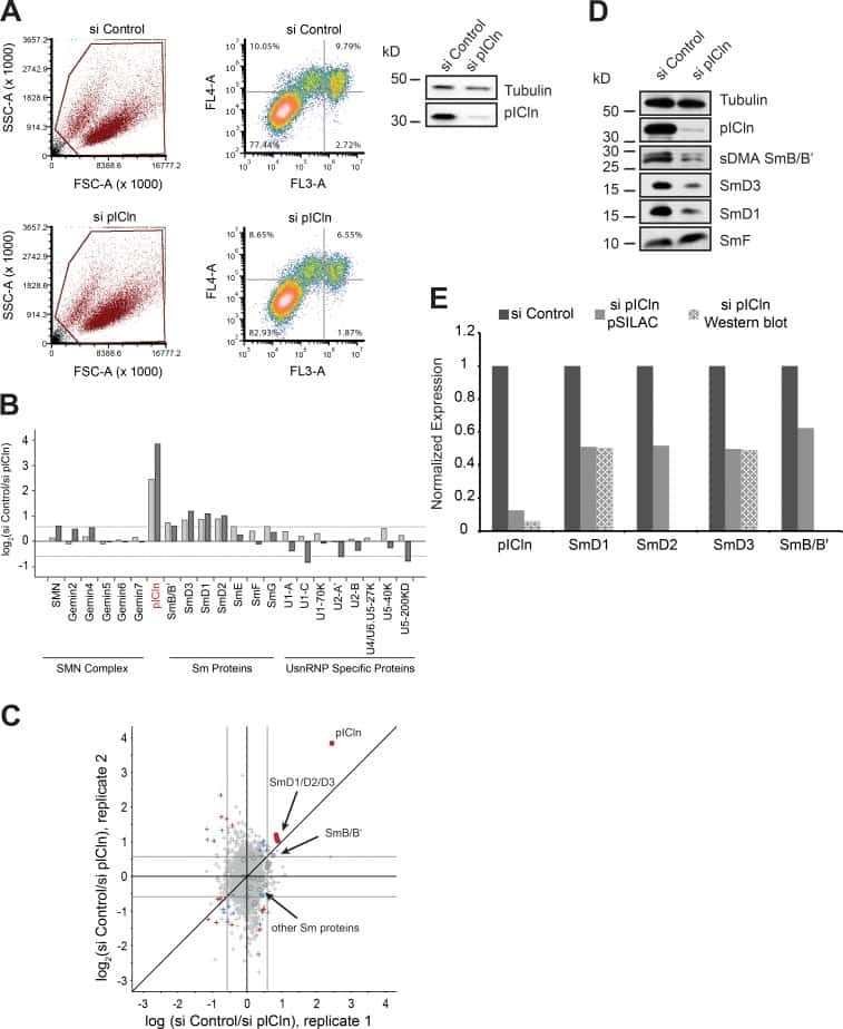
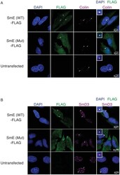
- Fig 3 NSM mutation causes mis-localization of the SNRPE/SmE protein. (A-B), Indirect immunofluorescence and confocal microscopy of HeLa cells transfected with either FLAG-tagged wild type or mutant SmE (WT/Mut) or left untransfected (negative control). Empty white arrowheads indicate localization pattern observed and filled white arrowheads indicate zoomed in region shown in the overly inset. (A), Top panel shows clear co-localization of wild type SmE (green) and coilin (magenta) in CBs and middle panel shows most of the mutant SmE (green) distributed in the cytoplasm with a minor fraction in the nucleus and co-localizing with coilin (magenta) in CBs. (B), While wild type SmE (green, top panel) co-localizes with SmD3 (magenta) in CBs and splicing speckles, the mutant SmE (green, middle panel) is predominantly cytoplasmic with marginal co-localization with SmD3 in CBs or in nuclear speckles.