 Explore
 Validate
 Learn
Learn Western blot
Antibody data
- Antibody Data
- Antigen structure
- References [0]
- Comments [0]
- Validations
- Western blot [2]
- Immunohistochemistry [1]
- Flow cytometry [1]
Submit
Validation data
Reference
Comment
Report error
- Product number
- ANC-021-200UL - Provider product page

- Provider
- Invitrogen Antibodies
- Product name
- Lynx1 (extracellular) Polyclonal Antibody
- Antibody type
- Polyclonal
- Antigen
- Other
- Reactivity
- Human, Mouse, Rat
- Host
- Rabbit
- Isotype
- IgG
- Vial size
- 200 µL
- Concentration
- 0.8 mg/mL
- Storage
- -20° C, Avoid Freeze/Thaw Cycles
No comments: Submit comment
Supportive validation
- Submitted by
- Invitrogen Antibodies (provider)
- Main image

- Experimental details
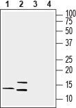
- Western blot analysis of rat brain lysate (lanes 1 and 3) and mouse brain lysate (lanes 2 and 4): - 1, 2. Anti-Lynx1 (extracellular) Antibody (#ANC-021), (1:200). 3, 4. Anti-Lynx1 (extracellular) Antibody , preincubated with Lynx1 (extracellular) Blocking Peptide (#BLP-NC021).
- Submitted by
- Invitrogen Antibodies (provider)
- Main image
- Experimental details
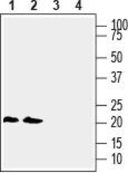
- Western blot analysis of human THP-1 monocytic leukemia cell line lysate (lanes 1 and 3) and human Jurkat T-cell leukemia cell line lysate (lanes 2 and 4): - 1, 2. Anti-Lynx1 (extracellular) Antibody (#ANC-021), (1:200). 3, 4. Anti-Lynx1 (extracellular) Antibody , preincubated with Lynx1 (extracellular) Blocking Peptide (#BLP-NC021).

Supportive validation
- Submitted by
- Invitrogen Antibodies (provider)
- Main image
- Experimental details
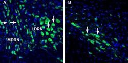
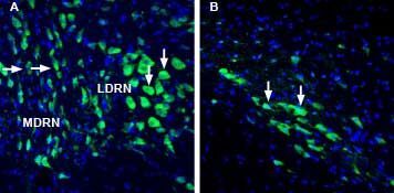
- Expression of Lynx1 in rat dorsal raphe nucleus and substantia nigra pars compacta - Immunohistochemical staining of perfusion-fixed frozen rat brain sections with Anti-Lynx1 (extracellular) Antibody (#ANC-021), (1:1000), followed by goat Anti-rabbit-AlexaFluor-488. A. Lynx1 staining in the dorsal raphe nucleus (DRN), (green) is detected in cells of the medial DRN (MDRN, horizontal arrows) and in cells of the lateral DRN (LDRN), (vertical arrows). B. Lynx1 immunoreactivity in the substantia nigra pars compacta (SNC), (green) is observed in cells of the SNC (arrows). Cell nuclei are stained with DAPI (blue).
Supportive validation
- Submitted by
- Invitrogen Antibodies (provider)
- Main image
- Experimental details
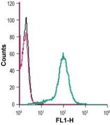
- Cell surface detection of Lynx1 in live intact human THP-1 monocytic leukemia cells: - (black line) cells. (red) Cells + goat- Anti-rabbit-FITC. (green) Cells + Anti-Lynx1 (extracellular) Antibody (#ANC-021), (2.5 µg) + goat- Anti-rabbit-FITC.
