Explore
 Validate
 Learn
Learn Immunohistochemistry
Antibody data
- Antibody Data
- Antigen structure
- References [0]
- Comments [0]
- Validations
- Immunohistochemistry [1]
Submit
Validation data
Reference
Comment
Report error
- Product number
- AF3940 - Provider product page

- Provider
- Novus Biologicals
- Product name
- Goat Polyclonal Tsukushi/TSK Antibody
- Antibody type
- Polyclonal
- Description
- Immunogen affinity purified. Detects human Tsukushi/TSK in direct ELISAs and Western blots. In direct ELISAs, approximately 40% cross-reactivity with recombinant mouse TSK is observed.
- Reactivity
- Human
- Host
- Goat
- Isotype
- IgG
- Vial size
- 100 ug
- Concentration
- LYOPH
- Storage
- Use a manual defrost freezer and avoid repeated freeze-thaw cycles. 12 months from date of receipt, -20 to -70 degreesC as supplied. 1 month, 2 to 8 degreesC under sterile conditions after reconstitution. 6 months, -20 to -70 degreesC under sterile conditions after reconstitution.
No comments: Submit comment
Supportive validation
- Submitted by
- Novus Biologicals (provider)
- Main image

- Experimental details
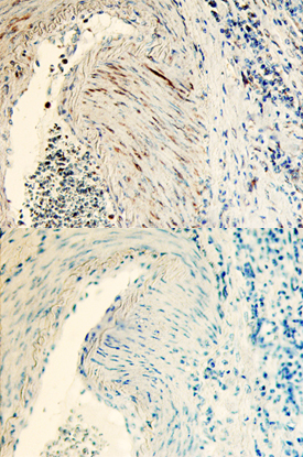
- Tsukushi/TSK in Human Colon Cancer Tissue. Tsukushi/TSK was detected in immersion fixed paraffin-embedded sections of human colon cancer tissue using Goat Anti-Human Tsukushi/TSK Antigen Affinity-purified Polyclonal Antibody (Catalog # AF3940) at 10 µg/mL overnight at 4 °C. Before incubation with the primary antibody, tissue was subjected to heat-induced epitope retrieval using Antigen Retrieval Reagent-Basic (Catalog # CTS013). Tissue was stained using the Anti-Goat HRP-DAB Cell & Tissue Staining Kit (brown; Catalog # CTS008) and counterstained with hematoxylin (blue). Lower panel shows a lack of labeling when primary antibodies are omitted and tissue is stained only with secondary antibody followed by incubation with detection reagents. Specific staining was localized to smooth muscle. View our protocol for Chromogenic IHC Staining of Paraffin-embedded Tissue Sections.