Explore
 Validate
 Learn
Learn Western blot
Antibody data
- Antibody Data
- Antigen structure
- References [1]
- Comments [0]
- Validations
- Western blot [3]
- Immunocytochemistry [3]
- Immunohistochemistry [4]
- Other assay [1]
Submit
Validation data
Reference
Comment
Report error
- Product number
- MA1-25043 - Provider product page

- Provider
- Invitrogen Antibodies
- Product name
- MAP2 Monoclonal Antibody (HM-2)
- Antibody type
- Monoclonal
- Antigen
- Other
- Description
- Recommended positive controls: rat brain. Store product as a concentrated solution. Centrifuge briefly prior to opening the vial.
- Reactivity
- Human, Mouse, Rat, Bovine, Chicken/Avian
- Host
- Mouse
- Isotype
- IgG
- Antibody clone number
- HM-2
- Vial size
- 50 µL
- Concentration
- 2.4 mg/mL
- Storage
- Store at 4°C short term. For long term storage, store at -20°C, avoiding freeze/thaw cycles.
Submitted references Brain clusterin protein isoforms and mitochondrial localization.
Herring SK, Moon HJ, Rawal P, Chhibber A, Zhao L
eLife 2019 Nov 18;8
eLife 2019 Nov 18;8
No comments: Submit comment
Supportive validation
- Submitted by
- Invitrogen Antibodies (provider)
- Main image
- Experimental details
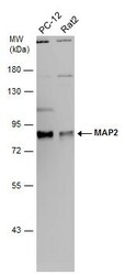
- Western Blot analysis of MAP2 was performed by separating 30 µg of various whole cell extracts by 7.5% SDS-PAGE. Proteins were transferred to a membrane and probed with a MAP2 Monoclonal Antibody (HM-2) (Product # MA1-25043) at a dilution of 1:2000.
- Submitted by
- Invitrogen Antibodies (provider)
- Main image
- Experimental details
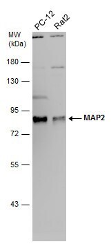
- Western Blot analysis of rat brain extract using MAP2 Monoclonal Antibody (HM-2) (Product # MA1-25043)at 1 µg/mL.

- Submitted by
- Invitrogen Antibodies (provider)
- Main image
- Experimental details
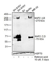
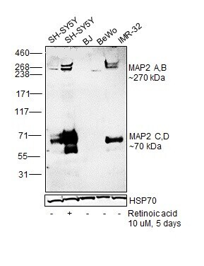
- Western blot was performed using Anti-MAP2 Monoclonal Antibody (HM-2) (Product # MA1-25043) and a ~270 kDa band corresponding to MAP2 A, B and ~70 kDa band corresponding to MAP2 C, D were observed across positive cell lines (SH-SY5Y and IMR-32); and the expression was increased in SH-SY5Y differentiated to neurons (10 µM Retinoic acid for 5 days). Whole cell extracts (30 µg lysate) of SH-SY5Y (Lane 1), SH-SY5Y treated with Retinoic acid (Lane 2), BJ (Lane 3), BeWo (Lane 4) and IMR-32 (Lane 5) were electrophoresed using NuPAGE™ 3-8% Tris-Acetate Protein Gel (Product # EA0378BOX). Resolved proteins were then transferred onto a Nitrocellulose membrane (Product # IB23001) by iBlot® 2 Dry Blotting System (Product # IB21001). The blot was probed with the primary antibody (2 µg/mL) and detected by chemiluminescence with Goat anti-Mouse IgG (H+L) Superclonal™ Recombinant Secondary Antibody, HRP (Product # A28177, 1:4,000 dilution) using the iBright FL 1000 (Product # A32752). Chemiluminescent detection was performed using SuperSignal™ West Dura Extended Duration Substrate (Product # 34076).
Supportive validation
- Submitted by
- Invitrogen Antibodies (provider)
- Main image
- Experimental details
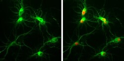
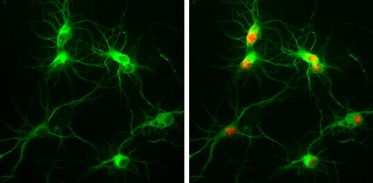
- Immunocytochemistry-Immunofluorescence analysis of MAP2 was performed in Rat E18 primary cortical neuron, DIV 8. Cells fixed in 4% paraformaldehyde at RT for 15 min. Green: MAP2 Monoclonal Antibody (HM-2) (Product # MA1-25043) diluted at 1:250. Red: NeuN, stained by NeuN antibody.
- Submitted by
- Invitrogen Antibodies (provider)
- Main image
- Experimental details
- Immunocytochemistry-Immunofluorescence analysis of MAP2 using MAP2 Monoclonal Antibody (HM-2) (Product # MA1-25043) at 2 µg/mL(red) with DAPI (blue). Cells were fixed and permeabilized with methanol followed by methanol: acetone.

- Submitted by
- Invitrogen Antibodies (provider)
- Main image
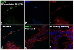
- Experimental details
- Immunofluorescence analysis of Microtubule-associated protein 2 was performed using 70% confluent log phase SH-SY5Y cells differentiated to neurons (Retinoic acid 10 uM for 5 days). The cells were fixed with 4% paraformaldehyde for 10 minutes, permeabilized with 0.1% Triton™ X-100 for 15 minutes, and blocked with 2% BSA for 45 minutes at room temperature. The cells were labeled with MAP2 Monoclonal Antibody (HM-2) (Product # MA1-25043) at 1:100 in 0.1% BSA, incubated at 4 degree celsius overnight and then labeled with Donkey anti-Mouse IgG (H+L) Highly Cross-Adsorbed Secondary Antibody, Alexa Fluor Plus 488 (Product # A32766), (1:2000 dilution), for 45 minutes at room temperature (Panel a: Green). Nuclei (Panel b: Blue) were stained with ProLong™ Diamond Antifade Mountant with DAPI (Product # P36962). F-actin (Panel c: Red) was stained with Rhodamine Phalloidin (Product # R415, 1:300 dilution). Panel d represents the merged image showing neuronal dendrites, cytoplasm, plasma membrane and cytoskeleton localization. Panel e represents undifferentiated SH-SY5Y cells. Panel f represents control cells with no primary antibody to assess background. The images were captured at 60X magnification.

Supportive validation
- Submitted by
- Invitrogen Antibodies (provider)
- Main image

- Experimental details
- Immunohistochemistry (Paraffin) analysis of MAP2 was performed in paraffin-embedded mouse brain tissue using MAP2 Monoclonal Antibody (HM-2) (Product # MA1-25043) at a dilution of 1:500.
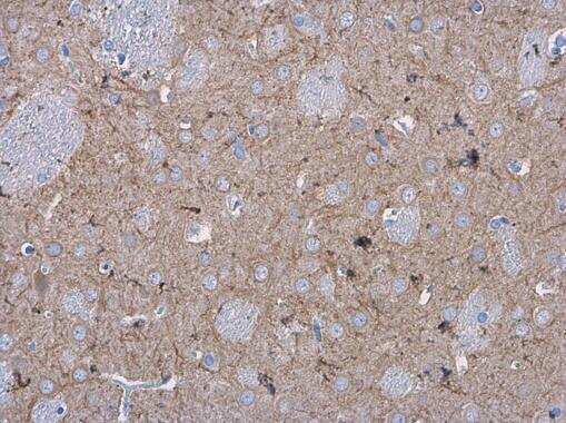
- Submitted by
- Invitrogen Antibodies (provider)
- Main image

- Experimental details
- Immunohistochemistry (Paraffin) analysis of MAP2 was performed in paraffin-embedded rat brain tissue using MAP2 Monoclonal Antibody (HM-2) (Product # MA1-25043) at a dilution of 1:500.
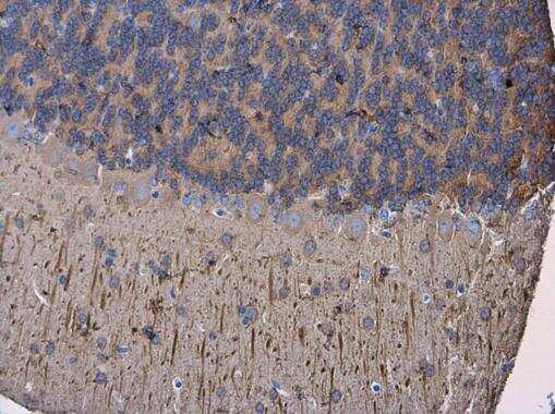
- Submitted by
- Invitrogen Antibodies (provider)
- Main image

- Experimental details
- Immunohistochemistry (Frozen) analysis of MAP2 was performed in frozen-sectioned adult mouse cerebellum tissue using MAP2 Monoclonal Antibody (HM-2) (Product # MA1-25043) at a dilution of 1:250 (Green). Blue: Fluoroshield with DAPI.

- Submitted by
- Invitrogen Antibodies (provider)
- Main image
- Experimental details
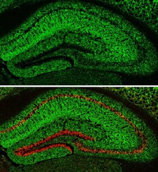
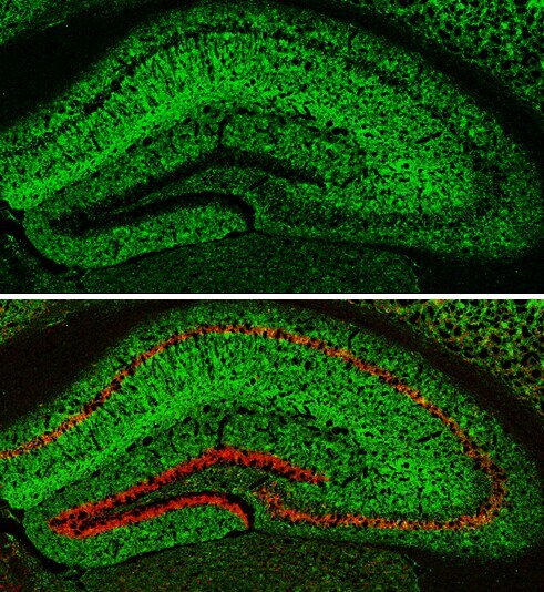
- Immunohistochemistry (Frozen) analysis of MAP2 was performed in frozen-sectioned adult mouse hippocampus tissue using MAP2 Monoclonal Antibody (HM-2) (Product # MA1-25043) at a dilution of 1:250 (Green). Red: NeuN, stained by NeuN antibody diluted at 1:500.
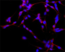
Supportive validation
- Submitted by
- Invitrogen Antibodies (provider)
- Main image
- Experimental details
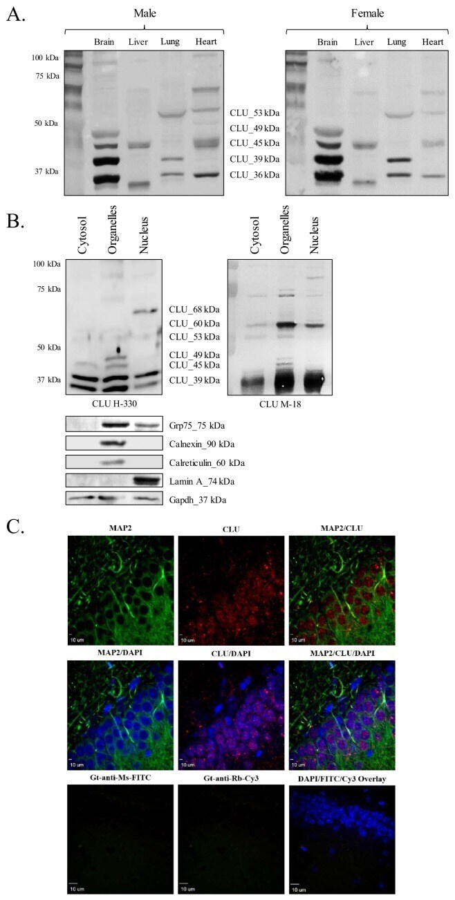
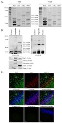
- Figure 1. CLU protein is expressed in the brain as multiple protein isoforms that have distinct cellular localizations. ( A ) Whole brain, liver, lung, and heart tissues were isolated from age-matched male (left panel) and female (right panel) wild-type (WT) mice and homogenized as indicated. Total protein was analyzed with SDS-PAGE and blots were probed for CLU immunoreactivity with anti-CLU H-330. ( B ) Cytosolic, organelle, and nuclear fractions were isolated from freshly harvested WT cortical tissue as indicated. Fractions were analyzed via SDS-PAGE and blots were probed for CLU immunoreactivity with anti-CLU H-330 (left panel) and anti-CLU M-18 (right panel). Blots were stripped and re-probed with fraction-/organelle-specific biochemical markers: Grp75 (mitochondria), calnexin and calreticulin (ER), lamin A (nucleus), and Gapdh (cytosol). ( C ) 40-uM-thick rodent brain sections were permeabilized and blocked as indicated and labeled with anti-MAP2 (green) or anti-GFAP ( Figure 1--figure supplement 1C ) and anti-CLU H-330 (red). Brain sections were then washed and probed with anti-mouse FITC (for MAP2) or anti-rat Cy5 (for GFAP) and pre-adsorbed anti-rabbit Cy3 (for CLU). To generate a secondary antibody control (bottom panel), one group of free-floating brain sections was incubated overnight in the same conditions without primary antibody. Brain sections from the hippocampal dentate gyrus were imaged at 4X ( Figure 1--figure supplement 1B ) and 40X using a customized