 Explore
 Validate
 Learn
Learn Western blot
Antibody data
- Antibody Data
- Antigen structure
- References [0]
- Comments [0]
- Validations
- Western blot [1]
- Immunocytochemistry [3]
Submit
Validation data
Reference
Comment
Report error
- Product number
- LS-C204540 - Provider product page

- Provider
- LSBio
- Product name
- MAP2 Antibody (clone 5H11) LS-C204540
- Antibody type
- Monoclonal
- Description
- Affinity purified
- Reactivity
- Human, Mouse, Rat, Bovine
- Host
- Mouse
- Isotype
- IgG
- Antibody clone number
- 5H11
- Storage
- Store at 4°C or -20°C. Avoid freeze-thaw cycles.
No comments: Submit comment
Supportive validation
- Submitted by
- LSBio (provider)
- Enhanced method
- Genetic validation
- Main image

- Experimental details
- Western blot of whole rat brain lysate probed with MAP2 / MAP-2 antibody antibody to MAP2. Note that the strong single band running at about 280Kda corresponds to MAP2.

Supportive validation
- Submitted by
- LSBio (provider)
- Enhanced method
- Genetic validation
- Main image

- Experimental details
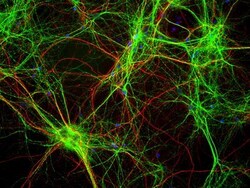
- Mixed neuron/glia cultures stained with MAP2 / MAP-2 antibody (green) and also rabbit antibody of neurofilament NF-H RPCA-NF-H (red). Since the NF-H protein is largely expressed in neuronal axons, while the MAP2 is only found in neuronal dendrites and perikarya, there is little overlap between these two staining patterns. DNA stain shows nuclei of neurons and non-neuronal cells (blue).

- Submitted by
- LSBio (provider)
- Main image

- Experimental details
- Mixed neuron/glia cultures stained with MAP2 / MAP-2 antibody (green) and also rabbit antibody of neurofilament NF-H RPCA-NF-H (red). Since the NF-H protein is largely expressed in neuronal axons, while the MAP2 is only found in neuronal dendrites and perikarya, there is little overlap between these two staining patterns. DNA stain shows nuclei of neurons and non-neuronal cells (blue).

- Submitted by
- LSBio (provider)
- Main image
- Experimental details
- Mixed neuron/glia cultures stained with MAP2 / MAP-2 antibody (green) and also rabbit antibody of neurofilament NF-H RPCA-NF-H (red). Since the NF-H protein is largely expressed in neuronal axons, while the MAP2 is only found in neuronal dendrites and perikarya, there is little overlap between these two staining patterns. DNA stain shows nuclei of neurons and non-neuronal cells (blue).
