Explore
 Validate
 Learn
Learn Western blot
Antibody data
- Antibody Data
- Antigen structure
- References [6]
- Comments [0]
- Validations
- Western blot [2]
- Immunocytochemistry [2]
- Immunohistochemistry [4]
Submit
Validation data
Reference
Comment
Report error
- Product number
- GTX11267 - Provider product page

- Provider
- GeneTex
- Proper citation
- GeneTex Cat#GTX11267, RRID:AB_369977
- Product name
- MAP2 antibody [HM-2]
- Antibody type
- Monoclonal
- Reactivity
- Human, Mouse, Rat, Bovine, Chicken/Avian
- Host
- Mouse
Submitted references An Inducible Alpha-Synuclein Expressing Neuronal Cell Line Model for Parkinson's Disease1.
α-Synuclein binds to the ER-mitochondria tethering protein VAPB to disrupt Ca2+ homeostasis and mitochondrial ATP production.
Histamine H3 receptor activation stimulates calcium mobilization in a subpopulation of rat striatal neurons in primary culture, but not in synaptosomes.
Astrocytic CCAAT/Enhancer Binding Protein δ Regulates Neuronal Viability and Spatial Learning Ability via miR-135a.
HIV-1-Tat Protein Inhibits SC35-mediated Tau Exon 10 Inclusion through Up-regulation of DYRK1A Kinase.
Persistent synaptic scaling independent of AMPA receptor subunit composition.
Vasquez V, Mitra J, Perry G, Rao KS, Hegde ML
Journal of Alzheimer's disease : JAD 2018;66(2):453-460
Journal of Alzheimer's disease : JAD 2018;66(2):453-460
α-Synuclein binds to the ER-mitochondria tethering protein VAPB to disrupt Ca2+ homeostasis and mitochondrial ATP production.
Paillusson S, Gomez-Suaga P, Stoica R, Little D, Gissen P, Devine MJ, Noble W, Hanger DP, Miller CCJ
Acta neuropathologica 2017 Jul;134(1):129-149
Acta neuropathologica 2017 Jul;134(1):129-149
Histamine H3 receptor activation stimulates calcium mobilization in a subpopulation of rat striatal neurons in primary culture, but not in synaptosomes.
Rivera-Ramírez N, Montejo-López W, López-Méndez MC, Guerrero-Hernández A, Molina-Hernández A, García-Hernández U, Arias-Montaño JA
Neurochemistry international 2016 Dec;101:38-47
Neurochemistry international 2016 Dec;101:38-47
Astrocytic CCAAT/Enhancer Binding Protein δ Regulates Neuronal Viability and Spatial Learning Ability via miR-135a.
Chu YY, Ko CY, Wang WJ, Wang SM, Gean PW, Kuo YM, Wang JM
Molecular neurobiology 2016 Aug;53(6):4173-4188
Molecular neurobiology 2016 Aug;53(6):4173-4188
HIV-1-Tat Protein Inhibits SC35-mediated Tau Exon 10 Inclusion through Up-regulation of DYRK1A Kinase.
Kadri F, Pacifici M, Wilk A, Parker-Struckhoff A, Del Valle L, Hauser KF, Knapp PE, Parsons C, Jeansonne D, Lassak A, Peruzzi F
The Journal of biological chemistry 2015 Dec 25;290(52):30931-46
The Journal of biological chemistry 2015 Dec 25;290(52):30931-46
Persistent synaptic scaling independent of AMPA receptor subunit composition.
Altimimi HF, Stellwagen D
The Journal of neuroscience : the official journal of the Society for Neuroscience 2013 Jul 17;33(29):11763-7
The Journal of neuroscience : the official journal of the Society for Neuroscience 2013 Jul 17;33(29):11763-7
No comments: Submit comment
Supportive validation
- Submitted by
- GeneTex (provider)
- Main image
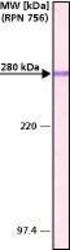
- Experimental details
- Rat brain extract (adult and newborn) was separated on SDS-PAGE and probed with Monoclonal PA To MAP2 (MO) CL:HM-2 (GTX11267). The antibody was developed with AlkPhos APA Mouse Fab ads HIgG and a NBTBCIP substrate. Antibody concentration: 1 μg/mL.
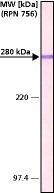
- Submitted by
- GeneTex (provider)
- Main image

- Experimental details
- Various whole cell extracts (30 ?g) were separated by 7.5% SDS-PAGE, and the membrane was blotted with MAP2 antibody [HM-2] (GTX11267) diluted at 1:2000. The signal was developed with Trident ECL plus-Enhanced.
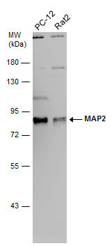
Supportive validation
- Submitted by
- GeneTex (provider)
- Main image

- Experimental details
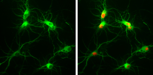
- MAP2 antibody [HM-2] detects MAP2 protein at by immunofluorescent analysis.Sample: Rat E18 primary cortical neuron, DIV 8. Cells were fixed in 4% paraformaldehyde at RT for 15 min.Green: MAP2 protein stained by MAP2 antibody [HM-2] (GTX11267) diluted at 1:250.Red: NeuN, stained by NeuN antibody (GTX132974) diluted at 1:250.
- Submitted by
- GeneTex (provider)
- Main image

- Experimental details
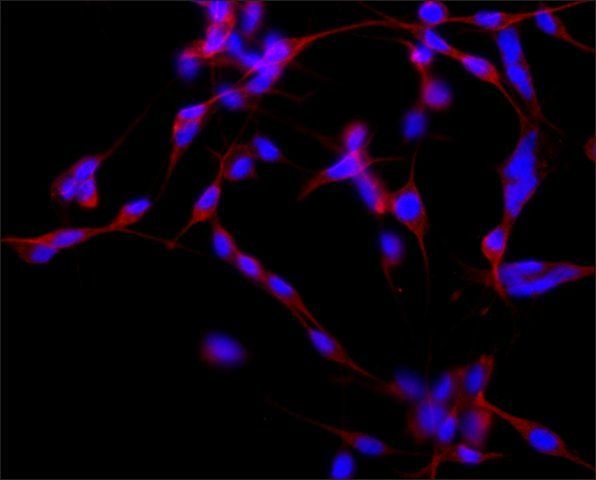
- ICC/IF analysis of B35 cells using MAP2 antibody [HM-2] at a concentration of 2 μg/ml (red) and DAPI (blue).
Supportive validation
- Submitted by
- GeneTex (provider)
- Main image

- Experimental details
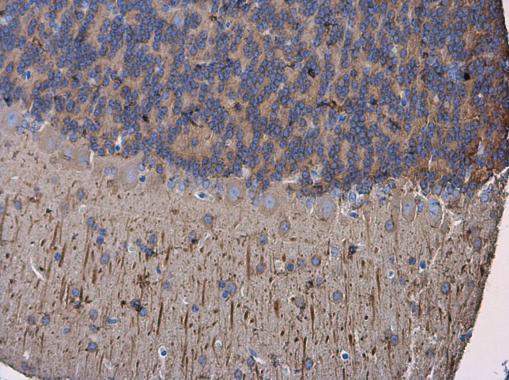
- MAP2 antibody [HM-2] detects MAP2 protein at cytoplasm in mouse brain by immunohistochemical analysis. Sample: Paraffin-embedded mouse brain. MAP2 antibody [HM-2] (GTX11267) diluted at 1:500.
- Submitted by
- GeneTex (provider)
- Main image
- Experimental details
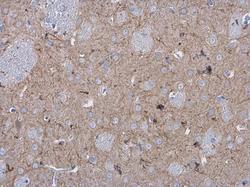
- MAP2 antibody [HM-2] detects MAP2 protein at cytoplasm in rat brain by immunohistochemical analysis. Sample: Paraffin-embedded rat brain. MAP2 antibody [HM-2] (GTX11267) diluted at 1:500.

- Submitted by
- GeneTex (provider)
- Main image

- Experimental details
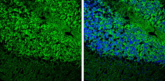
- MAP2 antibody [HM-2] detects MAP2 protein expression by immunohistochemical analysis.Sample: Frozen-sectioned adult mouse cerebellum. Green: MAP2 protein stained by MAP2 antibody [HM-2] (GTX11267) diluted at 1:250.Blue: Fluoroshield with DAPI (GTX30920).
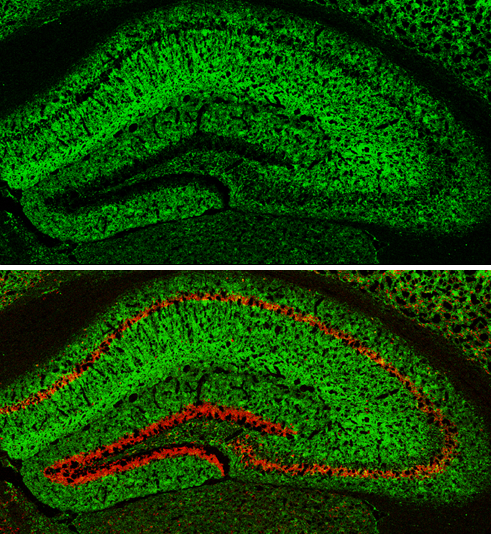
- Submitted by
- GeneTex (provider)
- Main image
- Experimental details
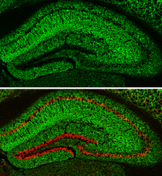
- MAP2 antibody [HM-2] detects MAP2 protein expression by immunohistochemical analysis.Sample: Frozen-sectioned adult mouse hippocampus. Green: MAP2 protein stained by MAP2 antibody [HM-2] (GTX11267) diluted at 1:250.Red: NeuN, stained by NeuN antibody (GTX132974) diluted at 1:500.