 Explore
 Validate
 Learn
Learn Western blot
 Immunocytochemistry
Antibody data
- Antibody Data
- Antigen structure
- References [31]
- Comments [0]
- Validations
- Immunocytochemistry [1]
- Immunohistochemistry [1]
- Other assay [5]
Submit
Validation data
Reference
Comment
Report error
- Product number
- MA5-12823 - Provider product page

- Provider
- Invitrogen Antibodies
- Product name
- MAP2 Monoclonal Antibody (AP20)
- Antibody type
- Monoclonal
- Antigen
- Other
- Description
- This antibody is specific to MAP2a and MAP2b, and will not cross-react with MAP1, MAP5, tubulin or tau. This clone reacts with dendrites and cell bodies of neurons but not with the neuronal processes. It does not react with the low MW form (70 kDa, MAP2c). The epitope has been mapped to aa 997-1332. The molecular weight of the doublet is ~280 kDa. For IHC staining, formalin-fixed tissues must be boiled in a 10mM citrate buffer, pH 6.0, for 10-20 min followed by cooling at room temperature for 20 min. Recommened positive controls include human glioblastoma T98G cells or brain tissue.
- Reactivity
- Human, Bovine
- Host
- Mouse
- Isotype
- IgG
- Antibody clone number
- AP20
- Vial size
- 500 µL
- Concentration
- 0.2 mg/mL
- Storage
- 4° C
Submitted references Modeling alpha-synuclein pathology in a human brain-chip to assess blood-brain barrier disruption.
Global transcriptome profile of the developmental principles of in vitro iPSC-to-motor neuron differentiation.
MeCP2 links heterochromatin condensates and neurodevelopmental disease.
Proteomic analysis of gemcitabine-resistant pancreatic cancer cells reveals that microtubule-associated protein 2 upregulation associates with taxane treatment.
Reduction of Inflammation and Enhancement of Motility after Pancreatic Islet Derived Stem Cell Transplantation Following Spinal Cord Injury.
Altering cortical input unmasks synaptic phenotypes in the YAC128 cortico-striatal co-culture model of Huntington disease.
Interleukin-1β secreted from betanodavirus-infected microglia caused the death of neurons in giant grouper brains.
Influence of cortical synaptic input on striatal neuronal dendritic arborization and sensitivity to excitotoxicity in corticostriatal coculture.
Abnormal serine phosphorylation of insulin receptor substrate 1 is associated with tau pathology in Alzheimer's disease and tauopathies.
Recovery of fertility in azoospermia rats after injection of adipose-tissue-derived mesenchymal stem cells: the sperm generation.
ABCA1 influences neuroinflammation and neuronal death.
Precocious and delayed neocortical synaptogenesis in fetal holoprosencephaly.
Hemimegalencephaly: foetal tauopathy with mTOR hyperactivation and neuronal lipidosis.
Hemimegalencephaly: foetal tauopathy with mTOR hyperactivation and neuronal lipidosis.
Reduction of lesion in injured rat spinal cord and partial functional recovery of motility after bone marrow derived mesenchymal stem cell transplantation.
Characterization of glioma stem cells through multiple stem cell markers and their specific sensitization to double-strand break-inducing agents by pharmacological inhibition of ataxia telangiectasia mutated protein.
CRH-R1 and CRH-R2 differentially modulate dendritic outgrowth of hippocampal neurons.
Phenotypic characteristics of hybrid cells generated by transferring neuronal nuclei into bone marrow stromal cell cytoplasts.
Mesenchymal stem cells improve the healing of ischemic colonic anastomoses (experimental study).
Optimizing orthotopic cell transplantation in the mouse adrenal gland.
eIF2alpha Phosphorylation-dependent translation in CA1 pyramidal cells impairs hippocampal memory consolidation without affecting general translation.
GPR56 is highly expressed in neural stem cells but downregulated during differentiation.
Characterization of neogenin-expressing neural progenitor populations and migrating neuroblasts in the embryonic mouse forebrain.
Architectural (Type IA) focal cortical dysplasia and parvalbumin immunostaining in temporal lobe epilepsy.
MKLP1 requires specific domains for its dendritic targeting.
Neural stem cells express non-neural markers during embryoid body coculture.
Isolation and characterization of neural stem cells from human fetal striatum.
The carboxy-terminal tail region of human Cav2.1 (P/Q-type) channel is not an essential determinant for its subcellular localization in cultured neurones.
BetaAPP and furin mRNA concentrates in immature senile plaques in the brain of Alzheimer patients.
Suppression of sprouting: An early function of NMDA receptors in the absence of AMPA/kainate receptor activity.
Suppression of sprouting: An early function of NMDA receptors in the absence of AMPA/kainate receptor activity.
Pediaditakis I, Kodella KR, Manatakis DV, Le CY, Hinojosa CD, Tien-Street W, Manolakos ES, Vekrellis K, Hamilton GA, Ewart L, Rubin LL, Karalis K
Nature communications 2021 Oct 8;12(1):5907
Nature communications 2021 Oct 8;12(1):5907
Global transcriptome profile of the developmental principles of in vitro iPSC-to-motor neuron differentiation.
Solomon E, Davis-Anderson K, Hovde B, Micheva-Viteva S, Harris JF, Twary S, Iyer R
BMC molecular and cell biology 2021 Feb 18;22(1):13
BMC molecular and cell biology 2021 Feb 18;22(1):13
MeCP2 links heterochromatin condensates and neurodevelopmental disease.
Li CH, Coffey EL, Dall'Agnese A, Hannett NM, Tang X, Henninger JE, Platt JM, Oksuz O, Zamudio AV, Afeyan LK, Schuijers J, Liu XS, Markoulaki S, Lungjangwa T, LeRoy G, Svoboda DS, Wogram E, Lee TI, Jaenisch R, Young RA
Nature 2020 Oct;586(7829):440-444
Nature 2020 Oct;586(7829):440-444
Proteomic analysis of gemcitabine-resistant pancreatic cancer cells reveals that microtubule-associated protein 2 upregulation associates with taxane treatment.
Le Large TYS, El Hassouni B, Funel N, Kok B, Piersma SR, Pham TV, Olive KP, Kazemier G, van Laarhoven HWM, Jimenez CR, Bijlsma MF, Giovannetti E
Therapeutic advances in medical oncology 2019;11:1758835919841233
Therapeutic advances in medical oncology 2019;11:1758835919841233
Reduction of Inflammation and Enhancement of Motility after Pancreatic Islet Derived Stem Cell Transplantation Following Spinal Cord Injury.
Karaoz E, Tepekoy F, Yilmaz I, Subasi C, Kabatas S
Journal of Korean Neurosurgical Society 2019 Mar;62(2):153-165
Journal of Korean Neurosurgical Society 2019 Mar;62(2):153-165
Altering cortical input unmasks synaptic phenotypes in the YAC128 cortico-striatal co-culture model of Huntington disease.
Schmidt ME, Buren C, Mackay JP, Cheung D, Dal Cengio L, Raymond LA, Hayden MR
BMC biology 2018 Jun 27;16(1):58
BMC biology 2018 Jun 27;16(1):58
Interleukin-1β secreted from betanodavirus-infected microglia caused the death of neurons in giant grouper brains.
Chiang YH, Wu YC, Chi SC
Developmental and comparative immunology 2017 May;70:19-26
Developmental and comparative immunology 2017 May;70:19-26
Influence of cortical synaptic input on striatal neuronal dendritic arborization and sensitivity to excitotoxicity in corticostriatal coculture.
Buren C, Tu G, Parsons MP, Sepers MD, Raymond LA
Journal of neurophysiology 2016 Aug 1;116(2):380-90
Journal of neurophysiology 2016 Aug 1;116(2):380-90
Abnormal serine phosphorylation of insulin receptor substrate 1 is associated with tau pathology in Alzheimer's disease and tauopathies.
Yarchoan M, Toledo JB, Lee EB, Arvanitakis Z, Kazi H, Han LY, Louneva N, Lee VM, Kim SF, Trojanowski JQ, Arnold SE
Acta neuropathologica 2014 Nov;128(5):679-89
Acta neuropathologica 2014 Nov;128(5):679-89
Recovery of fertility in azoospermia rats after injection of adipose-tissue-derived mesenchymal stem cells: the sperm generation.
Cakici C, Buyrukcu B, Duruksu G, Haliloglu AH, Aksoy A, Isık A, Uludag O, Ustun H, Subası C, Karaoz E
BioMed research international 2013;2013:529589
BioMed research international 2013;2013:529589
ABCA1 influences neuroinflammation and neuronal death.
Karasinska JM, de Haan W, Franciosi S, Ruddle P, Fan J, Kruit JK, Stukas S, Lütjohann D, Gutmann DH, Wellington CL, Hayden MR
Neurobiology of disease 2013 Jun;54:445-55
Neurobiology of disease 2013 Jun;54:445-55
Precocious and delayed neocortical synaptogenesis in fetal holoprosencephaly.
Sarnat HB, Flores-Sarnat L
Clinical neuropathology 2013 Jul-Aug;32(4):255-68
Clinical neuropathology 2013 Jul-Aug;32(4):255-68
Hemimegalencephaly: foetal tauopathy with mTOR hyperactivation and neuronal lipidosis.
Sarnat H, Flores-Sarnat L, Crino P, Hader W, Bello-Espinosa L
Folia neuropathologica 2012;50(4):330-45
Folia neuropathologica 2012;50(4):330-45
Hemimegalencephaly: foetal tauopathy with mTOR hyperactivation and neuronal lipidosis.
Sarnat H, Flores-Sarnat L, Crino P, Hader W, Bello-Espinosa L
Folia neuropathologica 2012;50(4):330-45
Folia neuropathologica 2012;50(4):330-45
Reduction of lesion in injured rat spinal cord and partial functional recovery of motility after bone marrow derived mesenchymal stem cell transplantation.
Karaoz E, Kabatas S, Duruksu G, Okcu A, Subasi C, Ay B, Musluman M, Civelek E
Turkish neurosurgery 2012;22(2):207-17
Turkish neurosurgery 2012;22(2):207-17
Characterization of glioma stem cells through multiple stem cell markers and their specific sensitization to double-strand break-inducing agents by pharmacological inhibition of ataxia telangiectasia mutated protein.
Raso A, Vecchio D, Cappelli E, Ropolo M, Poggi A, Nozza P, Biassoni R, Mascelli S, Capra V, Kalfas F, Severi P, Frosina G
Brain pathology (Zurich, Switzerland) 2012 Sep;22(5):677-88
Brain pathology (Zurich, Switzerland) 2012 Sep;22(5):677-88
CRH-R1 and CRH-R2 differentially modulate dendritic outgrowth of hippocampal neurons.
Sheng H, Xu Y, Chen Y, Zhang Y, Xu X, He C, Ni X
Endocrine 2012 Jun;41(3):458-64
Endocrine 2012 Jun;41(3):458-64
Phenotypic characteristics of hybrid cells generated by transferring neuronal nuclei into bone marrow stromal cell cytoplasts.
Zhou Z, Xu Y, Zhong Q, Zheng J
Brain research bulletin 2012 Feb 10;87(2-3):303-11
Brain research bulletin 2012 Feb 10;87(2-3):303-11
Mesenchymal stem cells improve the healing of ischemic colonic anastomoses (experimental study).
Adas G, Arikan S, Karatepe O, Kemik O, Ayhan S, Karaoz E, Kamali G, Eryasar B, Ustek D
Langenbeck's archives of surgery 2011 Jan;396(1):115-26
Langenbeck's archives of surgery 2011 Jan;396(1):115-26
Optimizing orthotopic cell transplantation in the mouse adrenal gland.
Cardoso CC, Bornstein SR, Hornsby PJ
Cell transplantation 2010;19(5):565-72
Cell transplantation 2010;19(5):565-72
eIF2alpha Phosphorylation-dependent translation in CA1 pyramidal cells impairs hippocampal memory consolidation without affecting general translation.
Jiang Z, Belforte JE, Lu Y, Yabe Y, Pickel J, Smith CB, Je HS, Lu B, Nakazawa K
The Journal of neuroscience : the official journal of the Society for Neuroscience 2010 Feb 17;30(7):2582-94
The Journal of neuroscience : the official journal of the Society for Neuroscience 2010 Feb 17;30(7):2582-94
GPR56 is highly expressed in neural stem cells but downregulated during differentiation.
Bai Y, Du L, Shen L, Zhang Y, Zhang L
Neuroreport 2009 Jul 1;20(10):918-22
Neuroreport 2009 Jul 1;20(10):918-22
Characterization of neogenin-expressing neural progenitor populations and migrating neuroblasts in the embryonic mouse forebrain.
Fitzgerald DP, Cole SJ, Hammond A, Seaman C, Cooper HM
Neuroscience 2006 Oct 27;142(3):703-16
Neuroscience 2006 Oct 27;142(3):703-16
Architectural (Type IA) focal cortical dysplasia and parvalbumin immunostaining in temporal lobe epilepsy.
Garbelli R, Meroni A, Magnaghi G, Beolchi MS, Ferrario A, Tassi L, Bramerio M, Spreafico R
Epilepsia 2006 Jun;47(6):1074-8
Epilepsia 2006 Jun;47(6):1074-8
MKLP1 requires specific domains for its dendritic targeting.
Xu X, He C, Zhang Z, Chen Y
Journal of cell science 2006 Feb 1;119(Pt 3):452-8
Journal of cell science 2006 Feb 1;119(Pt 3):452-8
Neural stem cells express non-neural markers during embryoid body coculture.
Denham M, Huynh T, Dottori M, Allen G, Trounson A, Mollard R
Stem cells (Dayton, Ohio) 2006 Apr;24(4):918-27
Stem cells (Dayton, Ohio) 2006 Apr;24(4):918-27
Isolation and characterization of neural stem cells from human fetal striatum.
Li X, Xu J, Bai Y, Wang X, Dai X, Liu Y, Zhang J, Zou J, Shen L, Li L
Biochemical and biophysical research communications 2005 Jan 14;326(2):425-34
Biochemical and biophysical research communications 2005 Jan 14;326(2):425-34
The carboxy-terminal tail region of human Cav2.1 (P/Q-type) channel is not an essential determinant for its subcellular localization in cultured neurones.
Hu Q, Saegusa H, Hayashi Y, Tanabe T
Genes to cells : devoted to molecular & cellular mechanisms 2005 Feb;10(2):87-96
Genes to cells : devoted to molecular & cellular mechanisms 2005 Feb;10(2):87-96
BetaAPP and furin mRNA concentrates in immature senile plaques in the brain of Alzheimer patients.
Marcinkiewicz M
Journal of neuropathology and experimental neurology 2002 Sep;61(9):815-29
Journal of neuropathology and experimental neurology 2002 Sep;61(9):815-29
Suppression of sprouting: An early function of NMDA receptors in the absence of AMPA/kainate receptor activity.
Lin SY, Constantine-Paton M
The Journal of neuroscience : the official journal of the Society for Neuroscience 1998 May 15;18(10):3725-37
The Journal of neuroscience : the official journal of the Society for Neuroscience 1998 May 15;18(10):3725-37
No comments: Submit comment
Supportive validation
- Submitted by
- Invitrogen Antibodies (provider)
- Main image

- Experimental details
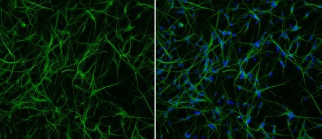
- Immunofluorescence analysis of neurons using anti-MAP2 antibody (Product # MA5-12823). GABAergic precursor cells derived from H9 embryonic stem cells were differentiated for 7 days and stained with antibodies against MAP2 (Product # MA5-12823 at 1:200) followed by Alexa Fluor 488 goat anti-rabbit (Product # A-21206, green). Nuclear DNA was stained with DAPI (blue) in the merged image (right panel).
Supportive validation
- Submitted by
- Invitrogen Antibodies (provider)
- Main image

- Experimental details
- Immunohistochemical analysis of MAP2 using a monoclonal antibody (Product # MA5-12823).

Supportive validation
- Submitted by
- Invitrogen Antibodies (provider)
- Main image

- Experimental details
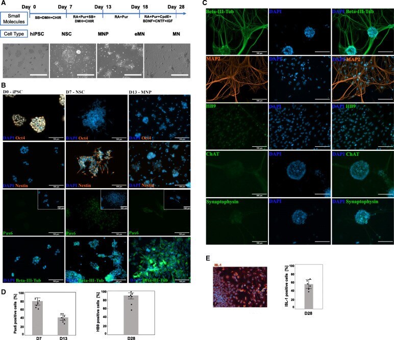
- Fig. 1 Differentiation of WTC-11 induced pluripotent stem cells (iPSC) into motor neurons (MN). a Schematic diagram of the overall experimental design. Shown are the small molecule stimuli for each developmental stage and the corresponding morphological changes in the course of iPSC conversion into MN. The scale bar for the phase contrast images is 400 mum; ( b ) Representative immunofluorescence images of key pluripotency (Oct4) and pan-neuronal (Nestin, Pax6, and beta-III tubulin) biomarkers indicated at efficient iPSC differentiation into neuronal stem cells (NSC) at day 7th post-induction (accumulation of Nestin is shown in orange). Upregulation of Pax6 and beta-III tubulin (shown in green) at day 13 of iPSC induction marked the formation of motor neuron progenitor (MNP) cells. c Immunostaining of neuronal structural proteins (beta-III-tubulin in green, MAP2 in orange), motor neuron specific markers (HB9 and ChAT in green), and synaptic vesicles protein (Synaptophysin in green) showed a pure population of mature MN at day 28 post-stimulation of iPSC. d Image quantification of Pax6- and HB9-labeled cells exemplified the percentage of cells that transitioned from NSC to MN. e Immunocytochemistry with anti-ISL1 antibody and quantitative analysis of biomarker positive cells at D28 of the differentiation protocol. The graphs show average of 6 cultures with > 300 cells in random fields for each culture. Scale bar: 100 mum
- Submitted by
- Invitrogen Antibodies (provider)
- Main image
- Experimental details
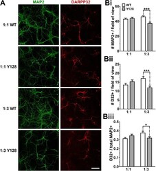
- Fig. 5 Neuronal survival is compromised in YAC128 1:3 CS co-cultures. DIV21 WT and YAC128 co-cultures were fixed at DIV21 and stained for MAP2 and DARPP32 (D32). ( a ) Sample fields of view at 20X objective (scale bar = 100 mum). The numbers of ( Bi ) MAP2+ and ( Bii ) DARPP32+ neurons per field of view are reduced in YAC128 1:3 co-cultures. ( Biii ) The proportion of DARPP32+ neurons (# DARPP32+ divided by # MAP2+) surviving at DIV21 is also significantly lower in YAC128 1:3 co-cultures [ n = 30 fields of view from three independent cultures; two-way ANOVA with Bonferroni post-hoc analysis; * p < 0.05, *** p < 0.001]

- Submitted by
- Invitrogen Antibodies (provider)
- Main image

- Experimental details
- Fig. 5 alphaSyn-induced caspase-3 activation and neuroinflammation. a Representative merged images showing immunostaining for microtubule-associated protein 2 (gray, MAP2), tyrosine hydroxylase (red, TH), and Cleaved Caspase-3 (green, CC3) in the brain channel at 6-days post-exposure to alphaSyn fibrils or alphaSyn monomers. Scale bars: 100 mum. b Quantitation of the number of CC3 and MAP2- or TH-positive neurons. Statistical analysis by two-way ANOVA with Tukey's multiple comparisons test (3-4 randomly selected different areas per chip, n = 3 independent chips/experimental group, ** P = 0.0033, **** P < 0.0001, NS not significant). Error bars represent mean +- SEM. Scale bars: 100 mum. c Immunostaining of the astrocyte marker glial fibrillary acidic protein (magenta, GFAP) demonstrated activation of astrocytes (white arrow) at day 6 post exposure to alphaSyn fibrils compared to monomeric alphaSyn. Scale bar, 100 mum. d Immunostaining of the microglial CD68 (red) demonstrated activation of microglia (white arrow) at day 6 post exposure to alphaSyn fibrils compared to monomeric alphaSyn. Scale bar, 100 mum. e The secreted levels of tumor necrosis factor-alpha (TNF-alpha) in the alphaSyn fibril model. Statistical analysis is two-sided unpaired t -test ( n = 6-7 independent chips, ** P = 0.0023). Error bars represent mean +- SEM. f The secreted levels of proinflammatory cytokine interleukin-6 (IL-6) in the alphaSyn fibril model. Statistical analysis is two-sided unpaired t -test

- Submitted by
- Invitrogen Antibodies (provider)
- Main image
- Experimental details
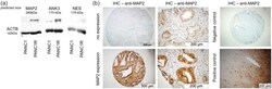
- Figure 3. Identification of predictive markers of gemcitabine resistance. (a) Western blot analysis of possible predictive biomarkers shows evident overexpression of MAP2 and ANK3 in PANC1R cells compared with the sensitive PANC1, in concordance with the MS/MS results. NES was expressed too low to be quantified properly. (b) Evaluation of MAP2 expression in TMAs. Representative images of TMA cores (4x) and images at higher magnification (40x) of a positive and negative staining of MAP2 in the glandular tumour islands. As a negative control (4x), slides without primary antibody was used. As a positive control (10x), human brain cortex was used. MAP2, microtubule-associated protein 2; MS/MS, tandem mass spectrometry; NES, Nestin; PANC1, gemcitabine-sensitive cell line; PANC1R, gemcitabine-resistant PANC1 cell line; TMA, tissue microarray.

- Submitted by
- Invitrogen Antibodies (provider)
- Main image

- Experimental details
- Figure 5. Immunofluorescence and immunohistochemistry of MAP2 in PANC1 and PANC1R cells and xenografts. (a) Immunofluorescence of PANC1 and PANC1R cells grown in chamber slides, and imaged at 100x magnification, showing a strong MAP2 (red)cytoplasmic staining in the PANC1R cells and low expression in PANC1, confirming our proteomic analysis. CK7 staining was performed as positive control (green), and DAPI was used to stain the nuclei (blue). This phenotype was conserved in our xenograft model as is shown by IHC (b), where MAP2 expression was overexpressed in the PANC1R tumours in vivo. DAPI, 4',6-diamidino-2-phenylindole; DFS, disease-free survival; IHC, immunohistochemistry; MAP2, microtubule-associated protein 2; OS, overall survival; PANC1, gemcitabine-sensitive cell line; PANC1R, gemcitabine-resistant PANC1 cell line.
